Biology:Ankyrin
| ANK1, erythrocytic | |
|---|---|
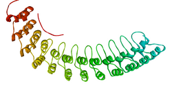
 Ribbon diagram of a fragment of the membrane-binding domain of ankyrin R.[1] | |
| Identifiers | |
| Symbol | ANK1 |
| Alt. symbols | AnkyrinR, Band2.1 |
| NCBI gene | 286 |
| HGNC | 492 |
| OMIM | 182900 |
| PDB | 1N11 |
| RefSeq | NM_000037 |
| UniProt | P16157 |
| Other data | |
| Locus | Chr. 8 p21.1-11.2 |
| Ankyrin repeat | |||||||||
|---|---|---|---|---|---|---|---|---|---|
| Identifiers | |||||||||
| Symbol | Ank | ||||||||
| Pfam | PF00023 | ||||||||
| InterPro | IPR002110 | ||||||||
| SMART | SM00248 | ||||||||
| PROSITE | PDOC50088 | ||||||||
| SCOP2 | 1awc / SCOPe / SUPFAM | ||||||||
| |||||||||
| ANK2, neuronal | |
|---|---|
| Identifiers | |
| Symbol | ANK2 |
| Alt. symbols | AnkyrinB |
| NCBI gene | 287 |
| HGNC | 493 |
| OMIM | 106410 |
| RefSeq | NM_001148 |
| UniProt | Q01484 |
| Other data | |
| Locus | Chr. 4 q25-q27 |
| ANK3, node of Ranvier | |
|---|---|
| Identifiers | |
| Symbol | ANK3 |
| Alt. symbols | AnkyrinG |
| NCBI gene | 288 |
| HGNC | 494 |
| OMIM | 600465 |
| RefSeq | NM_020987 |
| UniProt | Q12955 |
| Other data | |
| Locus | Chr. 10 q21 |
Ankyrins are a family of proteins that mediate the attachment of integral membrane proteins to the spectrin-actin based membrane cytoskeleton.[2] Ankyrins have binding sites for the beta subunit of spectrin and at least 12 families of integral membrane proteins. This linkage is required to maintain the integrity of the plasma membranes and to anchor specific ion channels, ion exchangers and ion transporters in the plasma membrane. The name is derived from the Greek word ἄγκυρα (ankyra) for "anchor".
Structure
Ankyrins contain four functional domains: an N-terminal domain that contains 24 tandem ankyrin repeats, a central domain that binds to spectrin, a death domain that binds to proteins involved in apoptosis, and a C-terminal regulatory domain that is highly variable between different ankyrin proteins.[2]
Membrane protein recognition
The 24 tandem ankyrin repeats are responsible for the recognition of a wide range of membrane proteins. These 24 repeats contain 3 structurally distinct binding sites ranging from repeat 1-14. These binding sites are quasi-independent of each other and can be used in combination. The interactions the sites use to bind to membrane proteins are non-specific and consist of: hydrogen bonding, hydrophobic interactions and electrostatic interactions. These non-specific interactions give ankyrin the property to recognise a large range of proteins as the sequence doesn't have to be conserved, just the properties of the amino acids. The quasi-independence means that if a binding site is not used, it won't have a large effect on the overall binding. These two properties in combination give rise to large repertoire of proteins ankyrin can recognise.
Subtypes
Ankyrins are encoded by three genes (ANK1, ANK2 and ANK3) in mammals. Each gene in turn produces multiple proteins through alternative splicing.
ANK1
The ANK1 gene encodes the AnkyrinR proteins. AnkyrinR was first characterized in human erythrocytes, where this ankyrin was referred to as erythrocyte ankyrin or band2.1.[3] AnkyrinR enables erythrocytes to resist shear forces experienced in the circulation. Individuals with reduced or defective ankyrinR have a form of hemolytic anemia termed hereditary spherocytosis.[4] In erythrocytes, AnkyrinR links the membrane skeleton to the Cl−/HCO3− anion exchanger.[5]
Ankyrin 1 links membrane receptor CD44 to the inositol triphosphate receptor and the cytoskeleton.[6]
It has been suggested that Ankyrin 1 interacts with KAHRP (shown via selective pull-downs, SPR and ELISA).[7]
ANK2

Subsequently, ankyrinB proteins (products of the ANK2 gene[8]) were identified in brain and muscle. AnkyrinB and AnkyrinG proteins are required for the polarized distribution of many membrane proteins including the Na+/K+ ATPase, the voltage gated Na+ channel and the Na+/Ca2+ exchanger.
ANK3
AnkyrinG proteins (products of the ANK3 gene[9]) were identified in epithelial cells and neurons. A large-scale genetic analysis conducted in 2008 shows the possibility that ANK3 is involved in bipolar disorder.[10][11]
See also
- DARPin (designed ankyrin repeat protein), an engineered antibody mimetic based on the structure of ankyrin repeats
References
- ↑ PDB: 1N11; "Crystal structure of a 12 ANK repeat stack from human ankyrinR". The EMBO Journal 21 (23): 6387–96. December 2002. doi:10.1093/emboj/cdf651. PMID 12456646.
- ↑ 2.0 2.1 "Spectrin and ankyrin-based pathways: metazoan inventions for integrating cells into tissues". Physiological Reviews 81 (3): 1353–92. July 2001. doi:10.1152/physrev.2001.81.3.1353. PMID 11427698. http://physrev.physiology.org/cgi/content/abstract/81/3/1353.
- ↑ "Identification and partial purification of ankyrin, the high affinity membrane attachment site for human erythrocyte spectrin". The Journal of Biological Chemistry 254 (7): 2533–41. April 1979. doi:10.1016/S0021-9258(17)30254-5. PMID 372182. http://www.jbc.org/cgi/pmidlookup?view=long&pmid=372182.
- ↑ "Hereditary spherocytosis associated with deletion of human erythrocyte ankyrin gene on chromosome 8". Nature 345 (6277): 736–9. June 1990. doi:10.1038/345736a0. PMID 2141669. Bibcode: 1990Natur.345..736L.
- ↑ "The membrane attachment protein for spectrin is associated with band 3 in human erythrocyte membranes". Nature 280 (5722): 468–73. August 1979. doi:10.1038/280468a0. PMID 379653. Bibcode: 1979Natur.280..468B.
- ↑ "CD44 interaction with ankyrin and IP3 receptor in lipid rafts promotes hyaluronan-mediated Ca2+ signaling leading to nitric oxide production and endothelial cell adhesion and proliferation". Experimental Cell Research 295 (1): 102–18. April 2004. doi:10.1016/j.yexcr.2003.12.025. PMID 15051494.
- ↑ "Interaction of Plasmodium falciparum knob-associated histidine-rich protein (KAHRP) with erythrocyte ankyrin R is required for its attachment to the erythrocyte membrane". Biochimica et Biophysica Acta (BBA) - Biomembranes 1838 (1 Pt B): 185–92. January 2014. doi:10.1016/j.bbamem.2013.09.014. PMID 24090929.
- ↑ "Mapping of a gene for long QT syndrome to chromosome 4q25-27". American Journal of Human Genetics 57 (5): 1114–22. November 1995. PMID 7485162.
- ↑ "Chromosomal localization of the ankyrinG gene (ANK3/Ank3) to human 10q21 and mouse 10". Genomics 27 (1): 189–91. May 1995. doi:10.1006/geno.1995.1023. PMID 7665168.
- ↑ "Collaborative genome-wide association analysis supports a role for ANK3 and CACNA1C in bipolar disorder". Nature Genetics 40 (9): 1056–8. September 2008. doi:10.1038/ng.209. PMID 18711365.
- ↑ "Channeling Mental Illness: GWAS Links Ion Channels, Bipolar Disorder". Schizophrenia Research Forum: News. schizophreniaforum.org. 2008-08-19. http://www.schizophreniaforum.org/new/detail.asp?id=1450.
External links
- Ankyrins at the US National Library of Medicine Medical Subject Headings (MeSH)
- Proteopedia 1n11 Ankyrin-R
it:Anchirina
 |
