Biology:Esophageal gland
| Esophageal glands | |
|---|---|
 Layers of Esophageal Wall: 1. Mucosa 2. Submucosa 3. Muscularis 4. Adventitia 5. Striated muscle 6. Striated and smooth 7. Smooth muscle 8. Lamina muscularis mucosae 9. Esophageal glands | |
 Section of the human esophagus. Moderately magnified. The section is transverse and from near the middle of the gullet. a. Fibrous covering. b. Divided fibers of longitudinal muscular coat. c. Transverse muscular fibers. d. Submucous or areolar layer. e. Muscularis mucosae. f. Mucous membrane, with vessels and part of a lymphoid nodule. g. Stratified epithelial lining. h. Mucous gland. i. Gland duct. m’. Striated muscular fibers cut across. | |
| Details | |
| Identifiers | |
| Latin | glandulae oesophageae |
| Anatomical terminology | |
The esophageal glands are glands that are part of the digestive system of various animals, including humans.
In humans
Esophageal glands in humans are a part of a human digestive system. They are a small compound racemose exocrine glands of the mucous type.[citation needed]
There are two types:
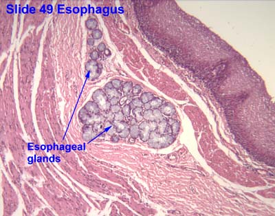
- Esophageal glands proper- mucous glands located in the submucosa. They are compound tubulo-alveolar glands. Some serous cells are present. These glands are more numerous in the upper third of the esophagus.[1] They secrete acid mucin for lubrication.[citation needed]
- Esophageal cardiac glands- mucous glands located near the cardiac orifice (esophago-gastric junction) in the lamina propria mucosae. They secrete neutral mucin[1] that protects the esophagus from acidic gastric juices. They are simple tubular or branched tubular glands.
- There are also mucous glands present at the pharyngo-esophageal junction in the lamina propria mucosae. These are simple tubular or branched tubular glands.[1]
Each opens upon the surface by a long excretory duct.[citation needed]
In monoplacophorans
Oesophageal gland is enlarged in large monoplacophoran species.[2]
In gastropods
Oesophageal gland or oesophageal pouch is a part of the digestive system of some gastropods. Oesophageal gland or pouch is a common feature in so-called basal gastropod clades, including Patelloidea, Vetigastropoda, Cocculiniformia, Neritimorpha and Neomphalina.[3]
The size of oesophageal gland of scaly-foot gastropod Chrysomallon squamiferum (family Peltospiridae within Neomphalina) is about two orders of magnitude over the usual size.[3] The scaly-foot gastropod houses endosymbiotic Bacteria in the oesophageal gland.[3] Chrysomallon squamiferum was thought to be the only species of Peltospiridae, that has enlarged oesophageal gland, but later it was shown that both species Gigantopelta has the oesophageal gland also enlarged.[4] In other peltospirids, the posterior portion of the oesophagus forms a pair of blind mid-oesophageal pouches or gutters extending only to the anterior end of the foot (Rhynchopelta, Peltospira, Nodopelta, Echinopelta, Pachydermia).[3] The same situation is in Melanodrymia within the family Melanodrymiidae.[3] Bathyphytophilidae and Lepetellidae are also known to have enlarged oesophageal pouches, however, though not to the extent of Chrysomallon.[3] Both are known to house endosymbiotic bacteria, in the case of bathyphytophilids most likely also in the oesophageal glands but in the lepetellids the endosymbionts are spread in the haemocoel.[3]
References
This article incorporates text in the public domain from the 20th edition of Gray's Anatomy (1918).
This article incorporates Creative Commons (CC-BY-4.0) text from the reference[3]
- ↑ 1.0 1.1 1.2 Nemeskeri, Agnes. Human Histology. Budapest: Apathy Istvan Foundation, Semmelweis University Budapest, Department of Human Morphology and Developmental Biology. pp. 16.
- ↑ Chen, Chong; Uematsu, Katsuyuki; Linse, Katrin; Sigwart, Julia D. (2017). "By more ways than one: Rapid convergence at hydrothermal vents shown by 3D anatomical reconstruction of Gigantopelta (Mollusca: Neomphalina)". BMC Evolutionary Biology 17 (1): 62. doi:10.1186/s12862-017-0917-z. ISSN 1471-2148. PMID 28249568.
- ↑ 3.0 3.1 3.2 3.3 3.4 3.5 3.6 3.7 Chen, C.; Copley, J.; Linse, K.; Rogers, A.; Sigwart, J. (2015). "The heart of a dragon: 3D anatomical reconstruction of the 'scaly-foot gastropod' (Mollusca: Gastropoda: Neomphalina) reveals its extraordinary circulatory system". Frontiers in Zoology 12: 13. doi:10.1186/s12983-015-0105-1. PMID 26085836. PMC 4470333. https://eprints.soton.ac.uk/378306/2/s12983-015-0105-1-s1.pdf.
- ↑ Chen, C.; Linse, K.; Roterman, C. N.; Copley, J. T.; Rogers, A. D. (2015). "A new genus of large hydrothermal vent‐endemic gastropod (Neomphalina: Peltospiridae)". Zoological Journal of the Linnean Society 175 (2): 319–335. doi:10.1111/zoj.12279.
External links
- Histology image: 49_07 at the University of Oklahoma Health Sciences Center
- Histology image: 10802loa – Histology Learning System at Boston University
 |  |

