Biology:External anal sphincter: Difference between revisions
(simplify) |
SpringEdit (talk | contribs) (update) |
||
| Line 13: | Line 13: | ||
| Nerve = Branch from the fourth sacral and contributions from the inferior hemorrhoidal branch of the [[Biology:Pudendal nerve|pudendal nerve]] | | Nerve = Branch from the fourth sacral and contributions from the inferior hemorrhoidal branch of the [[Biology:Pudendal nerve|pudendal nerve]] | ||
}} | }} | ||
The '''external anal sphincter''' (or '''sphincter ani externus''') is an oval tube of [[Biology:Skeletal muscle#Skeletal muscle cells|skeletal muscle fibers]].<ref name=":2242">{{Cite book |last=Standring |first=Susan |url=https://www.worldcat.org/oclc/1201341621 |title=Gray's Anatomy: The Anatomical Basis of Clinical Practice |year=1201 |isbn=978-0-7020-7707-4 |edition=42th |location=New York |pages=683 |oclc=1201341621}}</ref> Distally, it is adherent to the [[Biology:Integument|skin]] surrounding the margin of the [[Biology:Anus|anus]].'''<ref name=":02">{{Cite book |last=Gray |first=Henry |url=https://archive.org/details/anatomyofhumanbo1918gray/page/424/mode/2up?view=theater |title=Gray's Anatomy |year=1918 |edition=20th |pages=424–425}}</ref>''' It exhibits a resting state of tonical contraction | The '''external anal sphincter''' (or '''sphincter ani externus''') is an oval tube of [[Biology:Skeletal muscle#Skeletal muscle cells|skeletal muscle fibers]].<ref name=":2242">{{Cite book |last=Standring |first=Susan |url=https://www.worldcat.org/oclc/1201341621 |title=Gray's Anatomy: The Anatomical Basis of Clinical Practice |year=1201 |isbn=978-0-7020-7707-4 |edition=42th |location=New York |pages=683 |oclc=1201341621}}</ref> Distally, it is adherent to the [[Biology:Integument|skin]] surrounding the margin of the [[Biology:Anus|anus]].'''<ref name=":02">{{Cite book |last=Gray |first=Henry |url=https://archive.org/details/anatomyofhumanbo1918gray/page/424/mode/2up?view=theater |title=Gray's Anatomy |year=1918 |edition=20th |pages=424–425}}</ref>''' It exhibits a resting state of tonical contraction<ref name=":2242" /> and also contracts during the bulbospongiosus reflex.<ref name=Vodusek_2002>{{cite journal | vauthors = Vodušek DB, Deletis V | title = Intraoperative Neurophysiological Monitoring of the Sacral Nervous System | journal = Neurophysiology in Neurosurgery, A Modern Intraoperative Approach | pages = 153–165 | year = 2002 | doi = 10.1016/B978-012209036-3/50011-1 | isbn = 9780122090363 | s2cid = 78605592 }}</ref><ref name=Sarica_1987>{{cite journal | vauthors = Sarica Y, Karacan I | title = Bulbocavernosus reflex to somatic and visceral nerve stimulation in normal subjects and in diabetics with erectile impotence | journal = The Journal of Urology | volume = 138 | issue = 1 | pages = 55–58 | date = July 1987 | pmid = 3599220 | doi = 10.1016/S0022-5347(17)42987-9 }}</ref><ref name=Jiang_2009>{{cite journal | vauthors = Jiang XZ, Zhou CK, Guo LH, Chen J, Wang HQ, Zhang DQ, Shi BK, Xu ZS | display-authors = 6 | title = [Role of bulbocavernosus reflex to stimulation of prostatic urethra in pathologic mechanism of primary premature ejaculation] | language = zh | journal = Zhonghua Yi Xue Za Zhi | volume = 89 | issue = 46 | pages = 3249–3252 | date = December 2009 | pmid = 20193361 }}</ref><ref name=Podnar_2012>{{cite journal | vauthors = Podnar S | title = Clinical elicitation of the penilo-cavernosus reflex in circumcised men | journal = BJU International | volume = 109 | issue = 4 | pages = 582–585 | date = February 2012 | pmid = 21883821 | pmc = | doi = 10.1111/j.1464-410X.2011.10364.x | s2cid = 27143105 }}</ref> | ||
==Anatomy== | ==Anatomy== | ||
| Line 19: | Line 19: | ||
=== Structure === | === Structure === | ||
Historically, the sphincter was described as consisting of three parts (deep, superficial, and | Historically, the sphincter was described as consisting of three parts (deep, superficial, and subcutaneous). This is not supported by current anatomical knowledge. Some sources still describe it in two layers, deep (or proximal) and superficial (or distal or subcutaneous).<ref name=":2242" /> | ||
Some of the muscles fibres decussate at the anterior midline and posterior midline, so forming an anterior commissure and posterior commissure.<ref name=":2242" /> | Some of the muscles fibres decussate at the anterior midline and posterior midline, so forming an anterior commissure and posterior commissure.<ref name=":2242" /> | ||
| Line 47: | Line 47: | ||
==References== | ==References== | ||
{{Reflist}} | {{Reflist}} | ||
{{Gray's}} | |||
==External links== | ==External links== | ||
| Line 53: | Line 54: | ||
* {{NormanAnatomy|pelvis}} ({{NormanAnatomyFig|rectum}}) | * {{NormanAnatomy|pelvis}} ({{NormanAnatomyFig|rectum}}) | ||
{{Muscles of perineum}} | |||
{{Digestive tract}} | {{Digestive tract}} | ||
Latest revision as of 13:48, 2 May 2025
| External anal sphincter | |
|---|---|
 | |
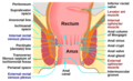
 Coronal section through the anal canal. B. Cavity of urinary bladder V.D. Ductus deferens. S.V. Seminal vesicle. R. Second part of rectum. A.C. Anal canal. L.A. Levator ani. I.S. Sphincter ani internus. E.S. Sphincter ani externus. | |
| Details | |
| Nerve | Branch from the fourth sacral and contributions from the inferior hemorrhoidal branch of the pudendal nerve |
| Actions | Keep the anal canal and orifice closed |
| Identifiers | |
| Latin | sphincter ani externus |
| Anatomical terms of muscle | |
The external anal sphincter (or sphincter ani externus) is an oval tube of skeletal muscle fibers.[1] Distally, it is adherent to the skin surrounding the margin of the anus.[2] It exhibits a resting state of tonical contraction[1] and also contracts during the bulbospongiosus reflex.[3][4][5][6]
Anatomy
The external anal sphincter is far more substantial than the internal anal sphincter. The proximal portion of external anal sphincter overlaps the internal anal sphincter (which terminates distally a little distance proximal to the anal orifice) superficially; where the two overlap, they are separated by the intervening conjoint longitudinal muscle.[1]
Structure
Historically, the sphincter was described as consisting of three parts (deep, superficial, and subcutaneous). This is not supported by current anatomical knowledge. Some sources still describe it in two layers, deep (or proximal) and superficial (or distal or subcutaneous).[1]
Some of the muscles fibres decussate at the anterior midline and posterior midline, so forming an anterior commissure and posterior commissure.[1]
Attachments
The muscle attaches anteriorly onto the perineal body, and posteriorly onto the anococcygeal ligament.[1]
Innervation
The sphincter receives innervation from the bilaterally paired inferior anal nerve (each a branch of the pudendal nerve which is derived from ventral rami of S2-S4). It may also receive additional motor innervation from the nerve to levator ani.[1]
Histology
The sphincter consists mostly of slow twitch fibers that allow extended continuous contraction.[1]
Gallery

Intestines
Anatomy of the human anus.
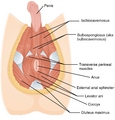
Muscles of male perineum.

Muscles of the female perineum.

Sagittal (vertical) section of bladder, penis, and urethra.


See also
- Internal anal sphincter
- Puborectalis muscle
References
- ↑ 1.0 1.1 1.2 1.3 1.4 1.5 1.6 1.7 Standring, Susan (1201). Gray's Anatomy: The Anatomical Basis of Clinical Practice (42th ed.). New York. pp. 683. ISBN 978-0-7020-7707-4. OCLC 1201341621. https://www.worldcat.org/oclc/1201341621.
- ↑ Gray, Henry (1918). Gray's Anatomy (20th ed.). pp. 424–425. https://archive.org/details/anatomyofhumanbo1918gray/page/424/mode/2up?view=theater.
- ↑ "Intraoperative Neurophysiological Monitoring of the Sacral Nervous System". Neurophysiology in Neurosurgery, A Modern Intraoperative Approach: 153–165. 2002. doi:10.1016/B978-012209036-3/50011-1. ISBN 9780122090363.
- ↑ "Bulbocavernosus reflex to somatic and visceral nerve stimulation in normal subjects and in diabetics with erectile impotence". The Journal of Urology 138 (1): 55–58. July 1987. doi:10.1016/S0022-5347(17)42987-9. PMID 3599220.
- ↑ "[Role of bulbocavernosus reflex to stimulation of prostatic urethra in pathologic mechanism of primary premature ejaculation]" (in zh). Zhonghua Yi Xue Za Zhi 89 (46): 3249–3252. December 2009. PMID 20193361.
- ↑ "Clinical elicitation of the penilo-cavernosus reflex in circumcised men". BJU International 109 (4): 582–585. February 2012. doi:10.1111/j.1464-410X.2011.10364.x. PMID 21883821.
This article incorporates text in the public domain from the 20th edition of Gray's Anatomy (1918)
External links
- Anatomy photo:42:13-0100 at the SUNY Downstate Medical Center - "The Male Perineum and the Penis: The External Anal Sphincter"
- perineum at The Anatomy Lesson by Wesley Norman (Georgetown University) (analtriangle3)
- pelvis at The Anatomy Lesson by Wesley Norman (Georgetown University) (rectum)
 |  |

