Biology:PSMA2
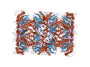
 Generic protein structure example |
Proteasome subunit alpha type-2 is a protein that in humans is encoded by the PSMA2 gene.[1][2][3] This protein is one of the 17 essential subunits (alpha subunits 1–7, constitutive beta subunits 1–7, and inducible subunits including beta1i, beta2i, beta5i) that contributes to the complete assembly of 20S proteasome complex.
Structure
Protein expression
The gene PSMA2 encodes a member of the peptidase T1A family, that is a 20S core alpha subunit.[3] Using FISH, the human gene HC3 (old nomenclature for PMSA2, 4.3kb with 3 exons) was mapped at chromosome band 6q27. The human protein proteasome subunit alpha type-2 is also known as 20S proteasome subunit alpha-2 (based on systematic nomenclature). The protein is 25.9 kDa in size and composed of 234 amino acids. The calculated theoretical pI of this protein is 6.77.[4]
Complex assembly
The proteasome is a multicatalytic proteinase complex with a highly ordered 20S core structure. This barrel-shaped core structure is composed of 4 axially stacked rings of 28 non-identical subunits: the two end rings are each formed by 7 alpha subunits, and the two central rings are each formed by 7 beta subunits. Three beta subunits (beta1, beta2, and beta5) each contains a proteolytic active site and has distinct substrate preferences. Proteasomes are distributed throughout eukaryotic cells at a high concentration and cleave peptides in an ATP/ubiquitin-dependent process in a non-lysosomal pathway.[5][6]
Function
Crystal structures of isolated 20S proteasome complex demonstrate that the two rings of beta subunits form a proteolytic chamber and maintain all their active sites of proteolysis within the chamber.[6] Concomitantly, the rings of alpha subunits form the entrance for substrates entering the proteolytic chamber. In an inactivated 20S proteasome complex, the gate into the internal proteolytic chamber are guarded by the N-terminal tails of specific alpha-subunit.[7][8] The proteolytic capacity of 20S core particle (CP) can be activated when CP associates with one or two regulatory particles (RP) on one or both side of alpha rings. These regulatory particles include 19S proteasome complexes, 11S proteasome complex, etc. Following the CP-RP association, the confirmation of certain alpha subunits will change and consequently cause the opening of substrate entrance gate. Besides RPs, the 20S proteasomes can also be effectively activated by other mild chemical treatments, such as exposure to low levels of sodium dodecylsulfate (SDS) or NP-14.[8][9] The eukaryotic proteasome recognized degradable proteins, including damaged proteins for protein quality control purpose or key regulatory protein components for dynamic biological precesses. An essential function of a modified proteasome, the immunoproteasome, is the processing of class I MHC peptides.
As a component of alpha ring, Proteasome subunit alpha type-2 contributes to the formation of heptameric alpha rings and substrate entrance gate. Importantly, alpha2 subunit plays a critical role in the assembly of 19S base and 20S. In a study using Saccharomyces cerevisiae proteasome core particle 20S and regulatory particle 19S (similar to human proteasome) base component to delineate the binding process between 19S and 20S, evidences showed that one 19S subunit, Rpt6, can insert its tail into the pocket formed by alpha2 and alpha3 subunit, facilitating the complex formation between 20S and 19S base component.[10]
Clinical significance
The proteasome and its subunits are of clinical significance for at least two reasons: (1) a compromised complex assembly or a dysfunctional proteasome can be associated with the underlying pathophysiology of specific diseases, and (2) they can be exploited as drug targets for therapeutic interventions. More recently, more effort has been made to consider the proteasome for the development of novel diagnostic markers and strategies. An improved and comprehensive understanding of the pathophysiology of the proteasome should lead to clinical applications in the future.
The proteasomes form a pivotal component for the ubiquitin–proteasome system (UPS) [11] and corresponding cellular Protein Quality Control (PQC). Protein ubiquitination and subsequent proteolysis and degradation by the proteasome are important mechanisms in the regulation of the cell cycle, cell growth and differentiation, gene transcription, signal transduction and apoptosis.[12] Subsequently, a compromised proteasome complex assembly and function lead to reduced proteolytic activities and the accumulation of damaged or misfolded protein species. Such protein accumulation may contribute to the pathogenesis and phenotypic characteristics in neurodegenerative diseases,[13][14] cardiovascular diseases,[15][16][17] inflammatory responses and autoimmune diseases,[18] and systemic DNA damage responses leading to malignancies.[19]
Several experimental and clinical studies have indicated that aberrations and deregulations of the UPS contribute to the pathogenesis of several neurodegenerative and myodegenerative disorders, including Alzheimer's disease,[20] Parkinson's disease[21] and Pick's disease,[22] Amyotrophic lateral sclerosis (ALS),[22] Huntington's disease,[21] Creutzfeldt–Jakob disease,[23] and motor neuron diseases, polyglutamine (PolyQ) diseases, Muscular dystrophies[24] and several rare forms of neurodegenerative diseases associated with dementia.[25] As part of the ubiquitin–proteasome system (UPS), the proteasome maintains cardiac protein homeostasis and thus plays a significant role in cardiac ischemic injury,[26] ventricular hypertrophy[27] and heart failure.[28] Additionally, evidence is accumulating that the UPS plays an essential role in malignant transformation. UPS proteolysis plays a major role in responses of cancer cells to stimulatory signals that are critical for the development of cancer. Accordingly, gene expression by degradation of transcription factors, such as p53, c-jun, c-Fos, NF-κB, c-Myc, HIF-1α, MATα2, STAT3, sterol-regulated element-binding proteins and androgen receptors are all controlled by the UPS and thus involved in the development of various malignancies.[29] Moreover, the UPS regulates the degradation of tumor suppressor gene products such as adenomatous polyposis coli (APC) in colorectal cancer, retinoblastoma (Rb). and von Hippel–Lindau tumor suppressor (VHL), as well as a number of proto-oncogenes (Raf, Myc, Myb, Rel, Src, Mos, Abl). The UPS is also involved in the regulation of inflammatory responses. This activity is usually attributed to the role of proteasomes in the activation of NF-κB which further regulates the expression of pro inflammatory cytokines such as TNF-α, IL-β, IL-8, adhesion molecules (ICAM-1, VCAM-1, P-selectin) and prostaglandins and nitric oxide (NO).[18] Additionally, the UPS also plays a role in inflammatory responses as regulators of leukocyte proliferation, mainly through proteolysis of cyclines and the degradation of CDK inhibitors.[30] Lastly, autoimmune disease patients with SLE, Sjögren syndrome and rheumatoid arthritis (RA) predominantly exhibit circulating proteasomes which can be applied as clinical biomarkers.[31]
References
- ↑ "Molecular cloning and sequence analysis of cDNAs for five major subunits of human proteasomes (multi-catalytic proteinase complexes)". Biochimica et Biophysica Acta (BBA) - Gene Structure and Expression 1089 (1): 95–102. May 1991. doi:10.1016/0167-4781(91)90090-9. PMID 2025653.
- ↑ "The primary structures of four subunits of the human, high-molecular-weight proteinase, macropain (proteasome), are distinct but homologous". Biochimica et Biophysica Acta (BBA) - Protein Structure and Molecular Enzymology 1079 (1): 29–38. August 1991. doi:10.1016/0167-4838(91)90020-Z. PMID 1888762.
- ↑ 3.0 3.1 "Entrez Gene: PSMA2 proteasome (prosome, macropain) subunit, alpha type, 2". https://www.ncbi.nlm.nih.gov/sites/entrez?Db=gene&Cmd=ShowDetailView&TermToSearch=5683.
- ↑ "IPC - Isoelectric Point Calculator". Biology Direct 11 (1): 55. October 2016. doi:10.1186/s13062-016-0159-9. PMID 27769290. PMC 5075173. http://isoelectric.ovh.org. Retrieved 2020-04-28.
- ↑ "Structure and functions of the 20S and 26S proteasomes". Annual Review of Biochemistry 65: 801–47. 1996. doi:10.1146/annurev.bi.65.070196.004101. PMID 8811196.
- ↑ 6.0 6.1 "Molecular architecture and assembly of the eukaryotic proteasome". Annual Review of Biochemistry 82: 415–45. 2013. doi:10.1146/annurev-biochem-060410-150257. PMID 23495936.
- ↑ "Structure of 20S proteasome from yeast at 2.4 A resolution". Nature 386 (6624): 463–71. April 1997. doi:10.1038/386463a0. PMID 9087403. Bibcode: 1997Natur.386..463G.
- ↑ 8.0 8.1 "A gated channel into the proteasome core particle". Nature Structural Biology 7 (11): 1062–7. November 2000. doi:10.1038/80992. PMID 11062564.
- ↑ "Regulation of murine cardiac 20S proteasomes: role of associating partners". Circulation Research 99 (4): 372–80. August 2006. doi:10.1161/01.RES.0000237389.40000.02. PMID 16857963.
- ↑ "Reconfiguration of the proteasome during chaperone-mediated assembly". Nature 497 (7450): 512–6. May 2013. doi:10.1038/nature12123. PMID 23644457. Bibcode: 2013Natur.497..512P.
- ↑ "Perilous journey: a tour of the ubiquitin–proteasome system". Trends in Cell Biology 24 (6): 352–9. June 2014. doi:10.1016/j.tcb.2013.12.003. PMID 24457024.
- ↑ "New insights into proteasome function: from archaebacteria to drug development". Chemistry & Biology 2 (8): 503–8. August 1995. doi:10.1016/1074-5521(95)90182-5. PMID 9383453.
- ↑ "The Ubiquitin–Proteasome System and Molecular Chaperone Deregulation in Alzheimer's Disease". Molecular Neurobiology 53 (2): 905–31. March 2016. doi:10.1007/s12035-014-9063-4. PMID 25561438.
- ↑ "Ubiquitin-proteasome system involvement in Huntington's disease". Frontiers in Molecular Neuroscience 7: 77. 2014. doi:10.3389/fnmol.2014.00077. PMID 25324717.
- ↑ "Proteotoxicity: an underappreciated pathology in cardiac disease". Journal of Molecular and Cellular Cardiology 71: 3–10. June 2014. doi:10.1016/j.yjmcc.2013.12.015. PMID 24380730.
- ↑ "Targeting the ubiquitin–proteasome system in heart disease: the basis for new therapeutic strategies". Antioxidants & Redox Signaling 21 (17): 2322–43. December 2014. doi:10.1089/ars.2013.5823. PMID 25133688.
- ↑ "Protein quality control and metabolism: bidirectional control in the heart". Cell Metabolism 21 (2): 215–26. February 2015. doi:10.1016/j.cmet.2015.01.016. PMID 25651176.
- ↑ 18.0 18.1 "The I kappa B kinase (IKK) and NF-kappa B: key elements of proinflammatory signalling". Seminars in Immunology 12 (1): 85–98. February 2000. doi:10.1006/smim.2000.0210. PMID 10723801.
- ↑ "Quality control mechanisms in cellular and systemic DNA damage responses". Ageing Research Reviews 23 (Pt A): 3–11. September 2015. doi:10.1016/j.arr.2014.12.009. PMID 25560147.
- ↑ "Role of the proteasome in Alzheimer's disease". Biochimica et Biophysica Acta (BBA) - Molecular Basis of Disease 1502 (1): 133–8. July 2000. doi:10.1016/s0925-4439(00)00039-9. PMID 10899438.
- ↑ 21.0 21.1 "The role of the ubiquitin-proteasomal pathway in Parkinson's disease and other neurodegenerative disorders". Trends in Neurosciences 24 (11 Suppl): S7–14. November 2001. doi:10.1016/s0166-2236(00)01998-6. PMID 11881748.
- ↑ 22.0 22.1 "Morphometrical reappraisal of motor neuron system of Pick's disease and amyotrophic lateral sclerosis with dementia". Acta Neuropathologica 104 (1): 21–8. July 2002. doi:10.1007/s00401-001-0513-5. PMID 12070660.
- ↑ "Marked increase in cerebrospinal fluid ubiquitin in Creutzfeldt-Jakob disease". Neuroscience Letters 139 (1): 47–9. May 1992. doi:10.1016/0304-3940(92)90854-z. PMID 1328965.
- ↑ "Limb-girdle muscular dystrophy". Current Neurology and Neuroscience Reports 3 (1): 78–85. January 2003. doi:10.1007/s11910-003-0042-9. PMID 12507416.
- ↑ "From neurodegeneration to neurohomeostasis: the role of ubiquitin". Drug News & Perspectives 16 (2): 103–8. March 2003. doi:10.1358/dnp.2003.16.2.829327. PMID 12792671.
- ↑ "The ubiquitin proteasome system and myocardial ischemia". American Journal of Physiology. Heart and Circulatory Physiology 304 (3): H337–49. February 2013. doi:10.1152/ajpheart.00604.2012. PMID 23220331.
- ↑ "Ubiquitin proteasome dysfunction in human hypertrophic and dilated cardiomyopathies". Circulation 121 (8): 997–1004. March 2010. doi:10.1161/CIRCULATIONAHA.109.904557. PMID 20159828.
- ↑ "The ubiquitin-proteasome system in cardiac physiology and pathology". American Journal of Physiology. Heart and Circulatory Physiology 291 (1): H1–H19. July 2006. doi:10.1152/ajpheart.00062.2006. PMID 16501026.
- ↑ "Potential for proteasome inhibition in the treatment of cancer". Drug Discovery Today 8 (7): 307–15. April 2003. doi:10.1016/s1359-6446(03)02647-3. PMID 12654543.
- ↑ "Regulatory functions of ubiquitination in the immune system". Nature Immunology 3 (1): 20–6. January 2002. doi:10.1038/ni0102-20. PMID 11753406.
- ↑ "Circulating proteasomes are markers of cell damage and immunologic activity in autoimmune diseases". The Journal of Rheumatology 29 (10): 2045–52. October 2002. PMID 12375310.
Further reading
- "Structure and functions of the 20S and 26S proteasomes". Annual Review of Biochemistry 65: 801–47. 1996. doi:10.1146/annurev.bi.65.070196.004101. PMID 8811196.
- "Death by deamination: a novel host restriction system for HIV-1". Cell 114 (3): 281–3. August 2003. doi:10.1016/S0092-8674(03)00602-0. PMID 12914693.
- "The genes for the alpha-type HC3 (PMSA2) and beta-type HC5 (PMSB1) subunits of human proteasomes map to chromosomes 6q27 and 7p12-p13 by fluorescence in situ hybridization". Genomics 27 (2): 377–9. May 1995. doi:10.1006/geno.1995.1062. PMID 7558012.
- "Human proteasome subunits from 2-dimensional gels identified by partial sequencing". Biochemical and Biophysical Research Communications 205 (3): 1785–9. December 1994. doi:10.1006/bbrc.1994.2876. PMID 7811265.
- "Isolation and characterization of alpha-type HC3 and beta-type HC5 subunit genes of human proteasomes". Journal of Molecular Biology 244 (1): 117–24. November 1994. doi:10.1006/jmbi.1994.1710. PMID 7966316.
- "Oligo-capping: a simple method to replace the cap structure of eukaryotic mRNAs with oligoribonucleotides". Gene 138 (1–2): 171–4. January 1994. doi:10.1016/0378-1119(94)90802-8. PMID 8125298.
- "Functional analysis of eukaryotic 20S proteasome nuclear localization signal". Experimental Cell Research 225 (1): 67–74. May 1996. doi:10.1006/excr.1996.0157. PMID 8635518.
- "HIV-1 tat inhibits the 20 S proteasome and its 11 S regulator-mediated activation". The Journal of Biological Chemistry 272 (13): 8145–8. March 1997. doi:10.1074/jbc.272.13.8145. PMID 9079628.
- "Construction and characterization of a full length-enriched and a 5'-end-enriched cDNA library". Gene 200 (1–2): 149–56. October 1997. doi:10.1016/S0378-1119(97)00411-3. PMID 9373149.
- "An endogenous inhibitor of human immunodeficiency virus in human lymphocytes is overcome by the viral Vif protein". Journal of Virology 72 (12): 10251–5. December 1998. doi:10.1128/JVI.72.12.10251-10255.1998. PMID 9811770.
- "Evidence for a newly discovered cellular anti-HIV-1 phenotype". Nature Medicine 4 (12): 1397–400. December 1998. doi:10.1038/3987. PMID 9846577.
- "The complete primary structure of mouse 20S proteasomes". Immunogenetics 49 (10): 835–42. September 1999. doi:10.1007/s002510050562. PMID 10436176.
- "Degradation of HIV-1 integrase by the N-end rule pathway". The Journal of Biological Chemistry 275 (38): 29749–53. September 2000. doi:10.1074/jbc.M004670200. PMID 10893419.
- "The hPLIC proteins may provide a link between the ubiquitination machinery and the proteasome". Molecular Cell 6 (2): 409–19. August 2000. doi:10.1016/S1097-2765(00)00040-X. PMID 10983987.
- "Isolation of a human gene that inhibits HIV-1 infection and is suppressed by the viral Vif protein". Nature 418 (6898): 646–50. August 2002. doi:10.1038/nature00939. PMID 12167863. Bibcode: 2002Natur.418..646S.
- "The RTP site shared by the HIV-1 Tat protein and the 11S regulator subunit alpha is crucial for their effects on proteasome function including antigen processing". Journal of Molecular Biology 323 (4): 771–82. November 2002. doi:10.1016/S0022-2836(02)00998-1. PMID 12419264.
- "Missense mutation in the tubulin-specific chaperone E (Tbce) gene in the mouse mutant progressive motor neuronopathy, a model of human motoneuron disease". The Journal of Cell Biology 159 (4): 563–9. November 2002. doi:10.1083/jcb.200208001. PMID 12446740.


