Biology:Fibronectin type II domain
| Fibronectin type II domain | |||||||||
|---|---|---|---|---|---|---|---|---|---|
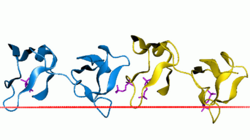
 Collagen-binding type II domain of seminal plasma protein PDC-109.[1] | |||||||||
| Identifiers | |||||||||
| Symbol | fn2 | ||||||||
| Pfam | PF00040 | ||||||||
| InterPro | IPR000562 | ||||||||
| SMART | SM00059 | ||||||||
| PROSITE | PDOC00022 | ||||||||
| SCOP2 | 1pdc / SCOPe / SUPFAM | ||||||||
| OPM superfamily | 115 | ||||||||
| OPM protein | 1h8p | ||||||||
| CDD | cd00062 | ||||||||
| |||||||||
Fibronectin type II domain is a collagen-binding protein domain. Fibronectin is a multi-domain glycoprotein, found in a soluble form in plasma, and in an insoluble form in loose connective tissue and basement membranes, that binds cell surfaces and various compounds including collagen, fibrin, heparin, DNA, and actin. Fibronectins are involved in a number of important functions e.g., wound healing; cell adhesion; blood coagulation; cell differentiation and migration; maintenance of the cellular cytoskeleton; and tumour metastasis.[2] The major part of the sequence of fibronectin consists of the repetition of three types of domains, which are called type I, II, and III.[3]
Type II domain is approximately sixty amino acids long,[4] contains four conserved cysteines involved in disulfide bonds and is part of the collagen-binding region of fibronectin. Type II domains occur two times in fibronectin. Type II domains have also been found in a range of proteins including blood coagulation factor XII; bovine seminal plasma proteins PDC-109 (BSP-A1/A2) and BSP-A3;[5] cation-independent mannose-6-phosphate receptor;[6] mannose receptor of macrophages;[7] 180 Kd secretory phospholipase A2 receptor;[8] DEC-205 receptor;[9] 72 Kd and 92 Kd type IV collagenase (EC 3.4.24.24);[10] and hepatocyte growth factor activator.[11]
Fibronectin type II domain and Lipid bilayer interaction
Fibronectin type II domain is part of the extracellular portions of EphA2 receptor proteins. FN2 domain on EphA2 receptors bears positively-charged components, namely K441 and R443, which attract and almost exclusively bind to anionic lipids such as anionic membrane lipid phosphatidylglycerol.[12] K441 and R443 together make up a membrane-binding motif that allows EphA2 receptors to attach to the cell membrane.[12]
Human proteins containing this domain
BSPH1; ELSPBP1; F12; FN1; HGFAC; IGF2R; LY75; MMP2; MMP9; MRC1; MRC1L1; MRC2; PLA2R1; SEL1L;
References
- ↑ "Sperm coating mechanism from the 1.8 A crystal structure of PDC-109-phosphorylcholine complex". Structure 10 (4): 505–14. April 2002. doi:10.1016/S0969-2126(02)00751-7. PMID 11937055.
- ↑ "Cloning and analysis of the promotor region of the human fibronectin gene". Proc. Natl. Acad. Sci. U.S.A. 84 (7): 1876–1880. 1987. doi:10.1073/pnas.84.7.1876. PMID 3031656.
- ↑ "Complete primary structure of bovine plasma fibronectin". Eur. J. Biochem. 161 (2): 441–453. 1986. doi:10.1111/j.1432-1033.1986.tb10464.x. PMID 3780752.
- ↑ "Fibronectin at a glance.". J Cell Sci 115 (20): 3861–3863. 2002. doi:10.1242/jcs.00059. PMID 12244123.
- ↑ "Complete amino acid sequence of BSP-A3 from bovine seminal plasma. Homology to PDC-109 and to the collagen-binding domain of fibronectin". Biochem. J. 243 (1): 195–203. 1987. doi:10.1042/bj2430195. PMID 3606570.
- ↑ Kornfeld S (1992). "Structure and function of the mannose 6-phosphate/insulinlike growth factor II receptors". Annu. Rev. Biochem. 61 (1): 307–330. doi:10.1146/annurev.bi.61.070192.001515. PMID 1323236.
- ↑ "Primary structure of the mannose receptor contains multiple motifs resembling carbohydrate-recognition domains". J. Biol. Chem. 265 (21): 12156–12162. 1990. doi:10.1016/S0021-9258(19)38325-5. PMID 2373685.
- ↑ "Cloning and expression of a membrane receptor for secretory phospholipases A2". J. Biol. Chem. 269 (3): 1575–1578. 1994. doi:10.1016/S0021-9258(17)42060-6. PMID 8294398.
- ↑ "The receptor DEC-205 expressed by dendritic cells and thymic epithelial cells is involved in antigen processing". Nature 375 (6527): 151–155. 1995. doi:10.1038/375151a0. PMID 7753172. Bibcode: 1995Natur.375..151J.
- ↑ "H-ras oncogene-transformed human bronchial epithelial cells (TBE-1) secrete a single metalloprotease capable of degrading basement membrane collagen". J. Biol. Chem. 263 (14): 6579–6587. 1988. doi:10.1016/S0021-9258(18)68680-6. PMID 2834383.
- ↑ "Molecular cloning and sequence analysis of the cDNA for a human serine protease responsible for activation of hepatocyte growth factor. Structural similarity of the protease precursor to blood coagulation factor XII". J. Biol. Chem. 268 (14): 10024–10028. 1993. doi:10.1016/S0021-9258(18)82167-6. PMID 7683665.
- ↑ 12.0 12.1 "Structures of the EphA2 Receptor at the Membrane: Role of Lipid Interactions". Structure 24 (2): 337–47. February 2016. doi:10.1016/j.str.2015.11.008. PMID 26724997.
External links
 |
