Biology:Marine biogenic calcification
Marine biogenic calcification refers to the production of calcium carbonate by organisms in the global ocean.
Marine biogenic calcification is the biologically mediated process by which marine organisms produce and deposit calcium carbonate minerals to form skeletal structures or hard tissues. This process is a fundamental aspect of the life cycle of some marine organisms, including corals, mollusks, foraminifera, certain types of plankton, and other calcifying marine invertebrates. The resulting structures, such as shells, skeletons, and coral reefs, function as protection, support, and shelter and create some of the most biodiverse habitats in the world. Marine biogenic calcifiers also play a key role in the biological carbon pump and the biogeochemical cycling of nutrients, alkalinity, and organic matter.[1]
Processes of Marine Biogenic Calcification
Biochemical mechanisms
Cellular and molecular processes of biogenic calcification
Calcium carbonate plays a fundamental role in the skeletal formation of marine calcifiers. The skeletal structures of these organisms are predominantly composed of calcium carbonate minerals, specifically aragonite and calcite. These structures provide support, protection, and housing for marine calcifiers and are formed through the biochemical processes of biomineralization to precipitate the crystal structures that form the hard tissues of these organisms.
The biogenic formation of calcium carbonate structures is the result of a combination of biological and physical processes such as genetics, cellular activity, crystal competition, growth in confined spaces, and self-organization processes. The composition of these structures, and the mechanisms involved in building them, are highly diverse. For example, some corals can incorporate both calcite and aragonite polymorphs into their skeletons. Some species, like corals and byrozoans, can incorporate other minerals to form complex protein matrices that perform specific functions.[2][3]
The key steps involved in marine biogenic calcification include the uptake of dissolved calcium ions (Ca2+) and carbonate ions (CO32-) from seawater, the precipitation of calcium carbonate crystals, and the controlled formation of skeletal structures through biomineralization processes. These organisms often regulate the calcification process through the secretion of organic molecules and proteins that influence the nucleation and growth of crystalline structures.
A range of biochemical calcification (biocalcification) mechanisms exist, indicated by the fact that marine calcifiers use several different forms of calcium carbonate minerals.[4] Within this range of mechanisms, there are two broad categories of biogenic calcification in marine organisms: extracellular mineralization and intracellular mineralization.[4] In particular, mollusks and corals use the extracellular strategy in which ion exchange pumps actively pump ions out of a cell into the extracellular space, where environmental conditions, such as pH, can be tightly controlled.[4] In contrast, during intracellular mineralization the calcium carbonate is formed within the organism and can either be kept within the organism as an internal structure or is later moved to the outside while retaining the cell membrane covering.[4] Broadly, the intracellular mechanism pumps ions into a vesicle within the cell.[4] This vesicle can then be secreted to the outside of the organism. Often, cells will fuse their membranes and combine these vesicles in order to build very large calcium carbonate structures that would not be possible within a single cell.[4]
Forms of calcium carbonate
The three most common calcium carbonate minerals are aragonite, calcite, and vaterite. Although these minerals have the same chemical formula (CaCO3), they are considered polymorphs because the atoms that make up the molecule are stacked in different arrangements. For example, aragonite minerals have an orthorhombic crystal lattice structure, while calcite crystals have a trigonal structure.[5] Some of the calcite polymorphs are further subdivided by relative magnesium content (Mg/Ca ratio), with calcite solubility increasing with increasing Mg.[6] The solubility of various forms of CaCO3differs in seawater; specifically, aragonite exhibits greater solubility compared to pure calcite, and the solubility of calcite rises with its magnesium content.[7]
Chemical processes and saturation state
The surface ocean engages in air-sea interactions and absorbs carbon dioxide (CO2) from the atmosphere, making the ocean the Earth's largest sink for atmospheric CO2. Carbon dioxide dissolves in and reacts with seawater to form carbonic acid. Subsequent reactions then produce carbonate (CO32−), bicarbonate (HCO3−), and hydrogen (H+) ions. Carbonate and bicarbonate are also deposited into the global ocean by rivers through the weathering of rock formations. The three species of carbon in seawater, carbon dioxide, bicarbonate, and carbonate, make up the total concentration of dissolved organic carbon (DIC) in the ocean. Approximately 90% of DIC is bicarbonate ions, 10% is carbonate ions, and <1% is dissolved carbon dioxide, with some spatial variation. The equilibria reactions between these species result in the buffering of seawater in terms of the concentrations of hydrogen ions present.[8]
The following chemical reactions exhibit the dissolution of carbon dioxide in seawater and its subsequent reaction with water:
CO2(g) + H2O(l) ⥨ H2CO3(aq)
H2CO3(aq) ⥨ HCO3-(aq) + H+(aq)
HCO3-(aq) ⥨ CO32−(aq) + H+(aq)
This series of reactions governs the pH levels in the ocean and also dictates the saturation state of seawater, indicating how saturated or unsaturated the seawater is with carbonate ions. Consequently, the saturation state significantly influences the balance between the dissolution and calcification processes in marine biogenic calcifiers. When seawater is oversaturated with calcium carbonate, the concentration of calcium ions and carbonate ions exceed the saturation point for a particular mineral, such as aragonite or calcite, which make up the skeletons of many marine organisms. Such conditions are favorable to marine calcifiers for the formation of calcium carbonate skeletons or shells. When seawater is undersaturated, meaning the concentration of calcium and carbonate ions is below the saturation point, it becomes challenging for marine calcifiers to build and maintain their skeletal structures, as the equilibrium conditions favor dissolution of calcium carbonate.[6] As a general rule, seawater that is undersaturated (Ω < 1) can dissolve the structures of calcifying organisms.[9] However, many organisms see negative effects on growth at saturation states above Ω = 1. For example, a saturation state of Ω = 3 is optimal for coral growth, so a saturation state Ω < 3 can potentially have negative effects on coral growth and survival.[10]
Calcium carbonate saturation can be determined using the following equation:
Ω = ([Ca2+][CO32−])/Ksp
where the numerator ([Ca2+][CO32−]) denotes the concentration of calcium and carbonate ions and the denominator (Ksp) refers to the mineral (solid) phase stoichiometric solubility product of calcium carbonate.[8]
When saturation is high, organisms can extract calcium and carbonate ions from seawater, forming solid crystals of calcium carbonate:
Ca2+(aq) + 2HCO3−(aq) → CaCO3(s) + CO2 + H2O
For marine calcifiers to build and maintain calcium carbonate structures, CaCO3 production must be greater than CaCO3 loss through physical, chemical, and biological processes. This net production can be thought of as follows:
CaCO3 accretion = CaCO3 production – CaCO3 dissolution – physical loss of CaCO3
The decreasing saturation of seawater with respect to calcium carbonate, associated with ocean acidification, a result of increased carbon dioxide (CO2) absorption by the oceans, poses a significant threat to marine calcifiers. As CO2 concentrations in seawater rise, a decrease in pH and a reduction in carbonate ion concentrations in seawater follows. This can make it difficult for marine organisms to precipitate and maintain their calcium carbonate structures, affecting growth, development, and overall health.[10][11]
The widespread use of calcification by marine organisms has relied on the ability of calcium carbonate to readily form in seawater, where the saturation states (Ω) of aragonite and calcite minerals have consistently surpassed Ω = 1 (indicating oversaturation) in surface waters for hundreds of millions of years.[12] The impacts of reduced calcium carbonate saturation on marine calcifiers have broader ecological implications, as these organisms play vital roles in marine ecosystems. For example, coral reefs, which are built by coral polyps secreting calcium carbonate skeletons, are particularly vulnerable to changes in calcium carbonate saturation.[13]
There is much debate in the scientific community on whether calcification rates correlate more with carbonate ions and saturation state or with pH. Some researchers state that a correlation exists between calcification and the Ω of carbonate ions in seawater.[14] Meanwhile, others state that from a physiological standpoint there are numerous marine organisms, and their calcification control is attributed more so to the concentrations of seawater bicarbonate (HCO3−) and protons (H+) rather than the Ω.[15] Further research is essential to gain a comprehensive understanding of the intricate connections between Ω, ocean acidification, and their impacts on the calcification rates of marine biogenic calcifiers, elucidating the distinct roles played by each.
Marine Calcifying Organisms
Corals
Coral reefs, physical structures formed from calcium carbonate, are important on biological and ecological scales to the regions they are endemic to.[11] Their robust calcification abilities have resulted in extensive calcium carbonate deposits, some housing significant hydrocarbon reserves.[16] However, this group only accounts for about 10% of the global production of calcium carbonate.[17]
Corals undergo extracellular calcification and first develop an organic matrix and skeleton on top of which they will form their calcite structures.[18] It is proposed that calcification via pH upregulation of the coral's extracellular calcifying fluid occurs at least in part via Ca2+-ATPase. Ca2+-ATPase is an enzyme in the calicoblastic epithelium that pumps Ca2+ ions into the calcifying region and ejects protons (H+).[19] This process circumvents the kinetic barriers to CaCO3precipitation that exist naturally in seawater.[19]
Molluscs
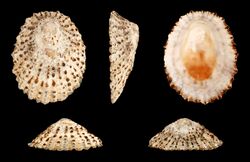
Mollusks are a diverse group including slugs, oysters, limpets, snails, scallops, mussels, clams, cephalopods and others. Mollusks employ a strategic approach to protect their soft tissues and deter predation by developing an external calcified shell. This process involves specialized cells following genetic instructions to synthesize minerals under non-equilibrium conditions.[20] The resulting minerals exhibit complex shapes and sizes along with being formed within a confined space.[20] These organisms also pump hydrogen out of the calcifying area so that it will not bond to the carbonate ions, which prevents crystallization of calcium carbonate.
Echinoderms
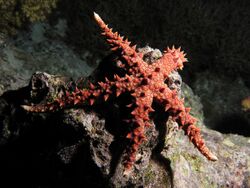
Echinoderms, of the phylum Echinodermata, include organisms such as sea stars, sea urchins, sand dollars, crinoids, sea cucumbers and brittle stars. These organisms form extensive endoskeletons consisting of magnesium-rich calcite.[21] Magnesium-rich calcite maintains the chemical composition of CaCO3, yet features substitutions of Mg for Ca as calcite and aragonite are mineral forms or polymorphs of CaCO3. Adult echinoderm skeletons consist of teeth, spines, tests, tubule feet, and in some cases, spicules.[22] Echinoderms serve as excellent blueprints for biomineralization. Adult sea urchins are a particularly popular species studied to better understand the molecular and cellular processes that the calcification and biomineralization of their skeletal structures requires.[21] Unlike many other marine calcifiers, echinoderm tests are not formed purely from calcite; instead, their structures also heavily consist of organic matrices that increases the toughness and strength of their endoskeletons.[22]
Crustaceans
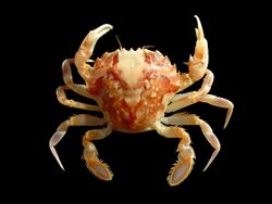
Crustaceans have a hard outer shell formed from calcium carbonate. These organisms form a network of chitin-protein fibers and then precipitate calcium carbonate within this matrix. The chitin-protein fibers are first hardened by sclerotization, or crosslinking of protein and polysaccharides, followed by the crosslinking of proteins with other proteins.[23] The presence of a hard, calcified exoskeleton means that the crustacean has to molt and shed the exoskeleton as its body size increases. This links molting cycles to calcification processes, making access to a regular source of calcium and carbonate ions crucial for the growth and survival of crustaceans.[23] Various body parts of the crustacean will have a different mineral content, varying the hardness at these locations with the harder areas being generally stronger. This calcite shell provides protection for the crustaceans, meaning between molting cycles the crustacean must avoid predators while it waits for its calcite shell to form and harden.
Foraminifera
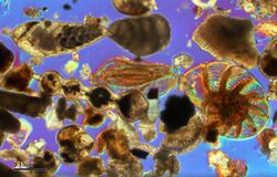
Foraminifera, or forams, are single-celled protists that form chambered shells (tests) from calcium carbonate.[24] Forams first appeared approximately 170 million years ago, and populate oceans globally. Forams are microscopic organisms, typically no larger than 1 mm in length.[24] The calcification and dissolution of their shells causes changes both in the surface seawater carbonate chemistry, and in deep-water chemistry.[24] These organisms are excellent paleo-proxies as they record ambient water chemistry during shell formation and are well-preserved in the sedimentary fossil record. Planktonic foraminifera, found in large numbers in the ocean, contribute significantly to oceanic carbonate production.[25] Unlike their benthic counterparts, more of these species have algal symbionts.
Coccolithophores
Phytoplankton, especially haptophytes such as coccolithophores, are also well known for their calcium carbonate production. It is estimated that these phytoplankton may contribute up to 70% of the global calcium carbonate precipitation, and coccolithophores are the largest phytoplankton contributors, along with diatoms and dinoflagellates.[26] Contributing between 1 and 10% of total ocean primary productivity, 200 species of coccolithophores live in the ocean, and under the right conditions they can form large blooms. These large bloom formations are a driving force for the export of calcium carbonate from the surface to the deep ocean in what is sometimes called “Coccolith rain”. As the coccolithophores sink to the seafloor they contribute to the vertical carbon dioxide gradient in the water column.[27]
Great Calcite Belt of the Southern Ocean
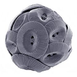
Coccolithophores produce calcite plates termed coccoliths which together cover the entire cell surface forming the coccosphere.[17] The coccoliths are formed using the intracellular strategy where the plates are formed in a coccoliths vesicle, but the product forming within the vesicle varies between the haploid and diploid phases. A coccolithophore in the haploid phase will produce what is called a holococcolith, while one in the diploid phase will produce heterococcoliths. Holococcoliths are small calcite crystals held together in an organic matrix, while heterococcoliths are arrays are larger, more complex calcite crystals.[17] These are often formed over a pre-existing template, giving each plate its particular structure and forming complex designs.[27] Each coccolithophore is a cell surrounded by the exoskeleton coccosphere, but there exists a wide range of sizes, shapes and architectures between different cells. Advantages of these plates may include protection against infection by viruses and bacteria, as well as protection from grazing zooplankton. The calcium carbonate exoskeleton enhances the amount of light the coccolithophore can uptake, increasing the level of photosynthesis. Finally, the coccoliths protect the phytoplankton from photodamage by UV light from the sun.[27]
The coccolithophores are also important in the geological history of Earth. The oldest coccolithophore fossil records are more than 209 million years old, placing their earliest presence in the Late Triassic period. Their calcium carbonate formation may have been the first deposition of carbonate on the seafloor.[27]
Corallinales (red algae)
Calcifying rhodophytes stock their filamentous cell walls with calcium carbonate and magnesium.[28] Corallinales is the one genus of red algae exists but their distribution ranges across the world's oceans.[29] Examples include Corallina, Neogoniolithon, and Harveylithon.[29] The magnesium-rich calcium carbonate of Corallinales cell wall provides shelter from predators and structural integrity in the intertidal zone.[30][31][32] The CaCO3 production in Coralline also plays a role in habitat formation and provides resources for benthic invertebrates.[31]
Calcifying bacteria
Evidence shows that some calcifying cyanobacteria strains have existed for millions of years and contributed to large land formations.[33] About 70 strains of cyanobacteria can precipitate calcium carbonate, including some strains of Synechococcus, Bacillus sphaericus, Bactilus subtilus,and Sporosarcina psychrophile.[34][31]
Morphological variations in calcium carbonate skeletons
Structural adaptations in different marine organisms
Diverse algae exhibit distinct mechanisms of CaCO3 formation, with calcification occurring internally or externally. Calcification may play a role in producing CO2 or supporting processes that need H+, based on the observed partial reaction.[35] Phytoplankton species relying on CO2 diffusion for photosynthesis may face limitations due to CO2 concentration and diffusion to the chloroplast's Rubisco site.[35] Calcifying macroalgae like Halimeda and Corallina also produce CaCO3 in alkaline extracellular spaces.[35]
Coccolithophorid phytoplankton form CaCO3 in crystalline structures known as coccoliths, with holococcoliths formed externally and heterococcoliths produced intracellularly.[36] Various coccolithophores produce two coccolith types: Heterococcoliths, from diploid cells, are complex, while holococcoliths, from haploid stages, are less studied. Factors influencing life cycle phase transitions and the role of specific proteins like GPA in coccolith morphology are explored.[36] Polysaccharides, particularly coccolith-associated polysaccharides (CAPs), emerge as key regulators of calcite growth and morphology. CAPs' diverse roles, including nucleation promotion and inhibition, vary between species.[36] External polysaccharides also influence coccolith adhesion and organization. Recent findings link cellular transport processes, carbonate saturation conditions, and regulatory processes determining calcite precipitation rate and morphology.[36] Unexpectedly, silicon's role in coccolith morphology regulation is species-dependent, highlighting physiological distinctions among coccolithophore groups. These revelations raise questions about ecological implications, evolutionary adaptations, and the impact of changing ocean silicate levels on coccolithogenesis.[36]
Calcification rates in coccolithophores often correlate with photosynthesis, implying a potential metabolic role.[35] Heterococcoliths develop inside intracellular vesicles, with coccolith formation showing a unity ratio with photosynthetic carbon fixation under high calcification rates. The variability in isotope fractionation and calcification mechanisms underscores these organisms' adaptability and complexity in responding to environmental factors.[35]
For corals, DIC from the seawater is absorbed and transferred to the coral skeleton. An anion exchanger will then be used to secrete DIC at the site of calcification. This DIC pool is also used by algal symbionts (dinoflagellates) that live in the coral tissue.[37] These algae photosynthesize and produce nutrients, some of which are passed to the coral. The coral in turn will emit ammonium waste products which the algae uptake as nutrients. There has been an observed tenfold increase in calcium carbonate formation in corals containing algal symbionts compared with corals that do not have this symbiotic relationship. The coral algal symbionts, Symbiodinium, show decreased populations with increased temperatures, often leaving the coral colorless and unable to photosynthesize and losing pigments (known as coral bleaching).[38]
Evolution of biogenic calcification
The evolution of biogenic calcification and carbonate structures within the eukaryotic domain is complex, highlighted by the distribution of mineralized skeletons across major clades.[39] Five out of the eight major clades feature species with mineralized skeletons, and all five clades involve organisms that precipitate calcite or aragonite.[39] Skeletal evolution occurred independently in foraminiferans and echinoderms, suggesting two separate origins of CaCO3 skeletons. The common ancestry for echinoderm and ascidian skeletons is less clear, but a conservative estimate indicates that carbonate skeletons evolved at least twenty-eight times within Euckarya.[39]
Phylogenetic insights highlight repeated innovations in carbonate skeleton evolution, raising questions about homology in underlying molecular processes.[39] Skeleton formation involves controlled mineral precipitation in specific biological environments, requiring directed calcium and carbonate transport, molecular templates, and growth inhibitors. Biochemical similarities, including the synthesis of acidic proteins and glycoproteins guiding mineralization, suggest an ancient capacity for carbonate formation in eukaryotes.[39] While skeletons may not share structural homology, underlying physiological pathways are common, reflecting multiple cooptations of molecular and physiological processes across eukaryotic organisms.[39]
The Cambrian Period marks a significant watershed in skeletal evolution, with the appearance of mineralized skeletons in various groups.[40] Skeletal diversity increased during this period, driven by predation pressure favoring protective armor evolution. The Cambrian radiation of mineralized skeletons was likely part of a broader animal diversity expansion.[40]
The evolution of mineralized skeletons during the Cambrian did not occur instantly, with a gradual increase in abundance and diversity over 25 million years.[41] Environmental changes and predation pressure played key roles in shaping skeletal evolution. The diversity of minerals and skeletal architectures during this period challenges explanations solely based on changing ocean chemistry. The interplay between genetic possibility and environmental opportunity, influenced by factors like increased oxygen tensions, likely contributed to Cambrian diversification.[41] Later Cambrian oceans witnessed a decline in mineralized skeletons, potentially influenced by high temperatures and pCO2 associated with a super greenhouse. Skeletal physiological responses to environmental conditions remain an area of study. Large-scale variations in carbonate chemistry suggest a connection between ocean chemistry and the mineralogy of carbonate precipitation.[39] Skeletal organisms that precipitate massive skeletons under limited physiological control show stratigraphic patterns corresponding to shifts in seawater chemistry.[42] This interplay between physiology, evolution, and environment underscores the complexity of mineralized skeleton evolution across geological time.
Calcium carbonate cycling and the biological carbon pump
The calcium carbonate cycle in the global ocean is of great significance to the biological, chemical, and physical state of the ocean. Mineral calcium carbonate most commonly presents as calcite in the ocean, and the majority of calcite is produced biologically in the upper layer of the ocean. CaCO3 material is exported from the upper ocean to sediments on the ocean floor where it either dissolves or is buried.[43] Alternatively, CaCO3 can dissolve or be remineralized within the water column prior to reaching the seafloor.
Upon reaching the seafloor, CaCO3 undergoes a diagenetic process that ends in either dissolution or burial.[44] The distribution of sediments consisting of calcium carbonate is fairly even across the global oceans, but specific locations are determined by the solubility and saturation level of calcium carbonate.[43]
The “biological carbon pump” is a colloquial term coined by scientists to summarize the global carbon cycle in the ocean and its relationship to the biological processes that occur throughout the ocean. The calcium carbonate cycle is inherently linked to the biological pump.[45] The formation of biogenic calcium carbonate by marine calcifiers is one way to add ballast to sinking particles and enhance transport of carbon to the deep ocean and seafloor.[45] The calcium carbonate counter pump refers to the biological process of precipitation of carbonate and the sinking of particulate inorganic carbon.[46] This process releases CO2 into the surface ocean and atmosphere across timescales spanning 100 to 1,000 years.[46] Its crucial role in regulating atmospheric pCO2 significantly influences global changes in atmospheric CO2 concentration.[47]
Inorganic sources of calcium carbonate
Of all the metals important to biogeochemical cycles in the ocean, calcium is one of the most significant in both its mobility and the role it plays in regulating climate over millions of years through its presence in calcium carbonate. Calcium has the ability to migrate relatively easily between the hydrosphere, the biosphere, and the crust of the Earth.[48]
Calcium and bicarbonate ions are largely deposited into the ocean from the weathering of rock formations and are transported via riverine input. This process occurs on very long timescales.[43] Weathering accounts for approximately 60-90% of solute calcium within the global calcium cycle.[48] Limestone rock, which consists mostly of calcite, is a prime example of a rich source of calcium to the ocean. The source of the majority of inorganic calcium present in the ocean is due to riverine deposition, though volcanic activity interacting with seawater does provide some calcium as well. The distribution of calcium sources described above is the case for both the present day oceanic calcium budget, and the historical budget over the last 25 million years.[49] The formation of biogenic calcium carbonate is the primary mechanism of removal of calcium in the ocean water column.[48]
Impact of environmental factors on calcification
Rising temperature and light exposure
Marine biogenic calcifiers, such as corals, are facing challenges due to increasing ocean temperatures, leading to prolonged warming events.[50] When sea surface temperatures exceed the local summer maximum monthly mean, coral bleaching and mortality occur as a result of the breakdown in symbiosis with Symbiodiniaceae.[50] Predicted increases in summer-time temperatures, coupled with ocean warming, are expected to impact coral health and overall rates of calcification, particularly in tropical regions where many corals already live close to their upper thermal limits.[50]
Corals are highly adapted to their local seasonal temperature and light conditions, influencing their physiology and calcification rates.[14] While increased temperature or light levels typically stimulate calcification up to a certain optimum, beyond which rates decline, the effects of temperature and light on the calcifying fluid chemistry are less clear.[14] Coral calcification is a biologically mediated process influenced by the regulation of internal calcifying fluid chemistry, including pH and dissolved inorganic carbon. The impacts of temperature and light on these factors remain a knowledge gap, with laboratory studies yielding contrasting results.[14] Decoupling the effects of temperature and light on calcification processes is challenging due to their seasonal co-variation, highlighting the need for further research to address this gap and enhance our understanding of how marine biogenic calcifiers respond to future climate change.[14]
Ocean Acidification
Calcifying organisms are particularly at risk due to changes in the chemical composition of ocean water associated with ocean acidification. As pH decreases due to ocean acidification, the availability of carbonate ions (CO32-) in seawater also decreases. Therefore, calcifying organisms experience difficulty building and maintaining their skeletons or shells in an acidic environment. There has been considerable debate in the literature regarding whether organisms are responding to reduced pH or reduced mineral saturation state as both variables decline with ocean acidification.[17][51][52] However, recent studies that have isolated the effects of saturation state independent of pH changes point toward saturation state as the most important factor impacting shell formation development.[50][53] However, we still need to fully constrain the carbonate chemistry to better interpret the ecological responses around ocean acidification.
Responses of marine calcifiers to reduced carbonate ion availability are seen in different ways. For example, coral reefs experience inhibited growth at decreased pH, and live calcium carbonate structures experience weakening of existing structures.[54][55] Other organisms are particularly vulnerable in the early stages of their life cycle. Bivalves for instance are particularly susceptible during early larval stages during initial shell formation since these early stages have a high energetic cost to the individual's development. In contrast, adult bivalves are considerably more resilient to reduced pH.[55]
Human interactions and applications
Economic importance
Shellfish industry
Ocean acidification (OA) presents a formidable threat to global shellfish production, particularly exerting its impact on calcification processes. Projections indicate that by the end of the century, mussel and oyster calcification could witness substantial reductions of 25% and 10%, respectively, as outlined in the IPCC IS92a scenario, which has an emissions trajectory that results in atmospheric CO2 reaching approximately 740 ppm in 2100.[56] These species, integral to coastal ecosystems and representing a significant portion of global aquaculture, play crucial roles as ecosystem engineers. The anticipated decline in calcification due to OA not only jeopardizes coastal biodiversity and ecosystem functioning but also carries the potential for considerable economic losses. For example, global aquaculture production for shellfish contributed US$29.2 billion to the world economy.[57]
Damaged shell surfaces, primarily resulting from reduced calcification rates, contribute to a significant decrease in sale prices, marking a critical economic concern. Economic assessments reveal that such damages, particularly impacting culture quasi-profits or applied cultural value, can lead to reductions ranging from 35% to 70%.[58] Furthermore, when accounting for assumed pH-driven changes occurring concurrently, quasi-profits diminish even more substantially, reaching levels of 49% to 84% across diverse OA scenarios. Consequently, the economic fallout is substantial, with the UK facing potential direct losses of £3 to £6 billion in GDP by 2100, and globally, costs exceeding US$100 billion.[59] These findings emphasize the urgent need for proactive measures to mitigate OA's impact on bivalve farming and underscore the importance of comprehensive climate policies to address these multifaceted challenges.
Coral reef tourism
For organisms relying on calcified structures (e.g. such as reef-associated organisms), OA can potentially disrupt entire ecosystems. As calcifiers play crucial roles in maintaining marine biodiversity, the repercussions of coral reef decline extend beyond economic considerations, emphasizing the urgency of comprehensive conservation efforts. Extensive degradation is occurring in the Caribbean and Western Atlantic region's coral reefs, stemming from issues like disease, overfishing, and a range of human activities.[60] Adding to the challenges, rapid climate-induced ocean warming and acidification exacerbate the threats to these vital ecosystems.[61] Tourism is integral to the Caribbean region with the sector contributing to over 15 percent of GDP and sustaining 13 percent of jobs in the region as a whole.[62] In the face of these challenges, the worldwide combined economic value of coral reefs is an estimated average of US$490 per hectare annually. Specific regions showcase the economic significance of coral reefs, with Hawai'i's contributing US$360 million annually to its economy, and the Philippine economy receiving at least US$1.06 billion each year from coral reefs.[60] In the St. Martin region, coral reefs contribute significantly, emphasizing the need for prioritized conservation and protection efforts.[60] Proposed solutions encompass ecological measures such as water quality management, sustainable fishing practices, ecological engineering, and marine spatial planning. Additionally, socio-economic strategies involve establishing a regional reef secretariat, integrating reef health into blue economy plans, and initiating a reef labeling program to foster corporate partnerships.[60]
See also
- Ocean acidification - a threat for marine biogenic calcification
- Protist shell
- Seashell
- Shell growth in estuaries
References
- ↑ Kawahata, Hodaka; Fujita, Kazuhiko; Iguchi, Akira; Inoue, Mayuri; Iwasaki, Shinya; Kuroyanagi, Azumi; Maeda, Ayumi; Manaka, Takuya et al. (2019-01-17). "Perspective on the response of marine calcifiers to global warming and ocean acidification—Behavior of corals and foraminifera in a high CO2 world "hot house"". Progress in Earth and Planetary Science 6 (1). doi:10.1186/s40645-018-0239-9. ISSN 2197-4284.
- ↑ Taylor, Paul D.; Lombardi, Chiara; Cocito, Silvia (November 2015). "Biomineralization in bryozoans: present, past and future" (in en). Biological Reviews 90 (4): 1118–1150. doi:10.1111/brv.12148. ISSN 1464-7931. PMID 25370313. https://onlinelibrary.wiley.com/doi/10.1111/brv.12148.
- ↑ Carter, Joseph G.; Harries, Peter; Malchus, Nikolaus; Sartori, Andre; Anderson, Laurie; Bieler, Rudiger; Bogan, Arthur; Coan, Eugene et al. (2012-02-01). "Treatise Online no. 48: Part N, Revised, Volume 1, Chapter 31: Illustrated Glossary of the Bivalvia". Treatise Online. doi:10.17161/to.v0i0.4322. ISSN 2153-4012. http://dx.doi.org/10.17161/to.v0i0.4322.
- ↑ 4.0 4.1 4.2 4.3 4.4 4.5 Plymouth Marine Laboratory. "The calcification process and measurement techniques" (PDF). International Atomic Energy Agency.
- ↑ Kleypas, Joan A. (2011), Hopley, David, ed. (in en), Ocean Acidification, Effects on Calcification, Encyclopedia of Earth Sciences Series, Dordrecht: Springer Netherlands, pp. 733–737, doi:10.1007/978-90-481-2639-2_118, ISBN 978-90-481-2639-2, https://doi.org/10.1007/978-90-481-2639-2_118, retrieved 2024-01-08
- ↑ 6.0 6.1 Ries, Justin B. (2011-07-15). "Skeletal mineralogy in a high-CO2 world". Journal of Experimental Marine Biology and Ecology 403 (1): 54–64. doi:10.1016/j.jembe.2011.04.006. ISSN 0022-0981. https://www.sciencedirect.com/science/article/pii/S002209811100164X.
- ↑ Andersson, AJ; Mackenzie, FT; Bates, NR (2008-12-23). "Life on the margin: implications of ocean acidification on Mg-calcite, high latitude and cold-water marine calcifiers". Marine Ecology Progress Series 373: 265–273. doi:10.3354/meps07639. ISSN 0171-8630. Bibcode: 2008MEPS..373..265A. http://dx.doi.org/10.3354/meps07639.
- ↑ 8.0 8.1 Dickson, Andrew. (2010). The carbon dioxide system in seawater: Equilibrium chemistry and measurements. Guide to Best Practices for Ocean Acidification Research and Data Reporting. 17-40.
- ↑ Azetsu-Scott, Kumiko; Clarke, Allyn; Falkner, Kelly; Hamilton, James; Jones, E. Peter; Lee, Craig; Petrie, Brian; Prinsenberg, Simon et al. (November 2010). "Calcium carbonate saturation states in the waters of the Canadian Arctic Archipelago and the Labrador Sea" (in en). Journal of Geophysical Research: Oceans 115 (C11). doi:10.1029/2009JC005917. ISSN 0148-0227. Bibcode: 2010JGRC..11511021A. https://agupubs.onlinelibrary.wiley.com/doi/10.1029/2009JC005917.
- ↑ 10.0 10.1 Yamamoto, S.; Kayanne, H.; Terai, M.; Watanabe, A.; Kato, K.; Negishi, A.; Nozaki, K. (2012-04-17). "Threshold of carbonate saturation state determined by CO<sub>2</sub> control experiment". Biogeosciences 9 (4): 1441–1450. doi:10.5194/bg-9-1441-2012. ISSN 1726-4189. Bibcode: 2012BGeo....9.1441Y.
- ↑ 11.0 11.1 Eyre, Bradley D.; Andersson, Andreas J.; Cyronak, Tyler (November 2014). "Benthic coral reef calcium carbonate dissolution in an acidifying ocean" (in en). Nature Climate Change 4 (11): 969–976. doi:10.1038/nclimate2380. ISSN 1758-6798. Bibcode: 2014NatCC...4..969E. https://www.nature.com/articles/nclimate2380.
- ↑ Yan, Xin; Zhang, Qi; Ma, Xinyue; Zhong, Yewen; Tang, Hengni; Mai, Sui (December 2023). "The mechanism of biomineralization: Progress in mineralization from intracellular generation to extracellular deposition" (in en). Japanese Dental Science Review 59: 181–190. doi:10.1016/j.jdsr.2023.06.005. PMID 37388714.
- ↑ Swart, P.K. "Coral Reefs: Canaries of the Sea, Rainforests of the Oceans". Nature Education Knowledge. 4 (3): 5.
- ↑ 14.0 14.1 14.2 14.3 14.4 Waldbusser, George G.; Hales, Burke; Langdon, Chris J.; Haley, Brian A.; Schrader, Paul; Brunner, Elizabeth L.; Gray, Matthew W.; Miller, Cale A. et al. (March 2015). "Saturation-state sensitivity of marine bivalve larvae to ocean acidification" (in en). Nature Climate Change 5 (3): 273–280. doi:10.1038/nclimate2479. ISSN 1758-6798. Bibcode: 2015NatCC...5..273W. https://www.nature.com/articles/nclimate2479.
- ↑ Cyronak, Tyler; Schulz, Kai G.; Jokiel, Paul L. (2015-12-13). "Response to Waldbusser et al. (2016): "Calcium carbonate saturation state: on myths and this or that stories"". ICES Journal of Marine Science 73 (3): 569–571. doi:10.1093/icesjms/fsv224. ISSN 1095-9289. https://doi.org/10.1093/icesjms/fsv224.
- ↑ Stanley, George D.; Fautin, Daphne G. (2001-03-09). "The Origins of Modern Corals" (in en). Science 291 (5510): 1913–1914. doi:10.1126/science.1056632. ISSN 0036-8075. PMID 11245198. https://www.science.org/doi/10.1126/science.1056632.
- ↑ 17.0 17.1 17.2 17.3 Zondervan, Ingrid; Zeebe, Richard E.; Rost, Björn; Riebesell, Ulf (June 2001). "Decreasing marine biogenic calcification: A negative feedback on rising atmospheric p CO 2" (in en). Global Biogeochemical Cycles 15 (2): 507–516. doi:10.1029/2000GB001321. ISSN 0886-6236. https://agupubs.onlinelibrary.wiley.com/doi/10.1029/2000GB001321.
- ↑ Kawaguti, Siro (1973-12-19). "Electron Microscopy on Symbiotic Algae in Reef Corals". Publications of the Seto Marine Biological Laboratory 20: 779–783. doi:10.5134/175743. ISSN 0037-2870. https://repository.kulib.kyoto-u.ac.jp/dspace/handle/2433/175743.
- ↑ 19.0 19.1 McCulloch, Malcolm T.; D’Olivo, Juan Pablo; Falter, James; Holcomb, Michael; Trotter, Julie A. (2017-05-30). "Coral calcification in a changing World and the interactive dynamics of pH and DIC upregulation" (in en). Nature Communications 8 (1): 15686. doi:10.1038/ncomms15686. ISSN 2041-1723. PMID 28555644. Bibcode: 2017NatCo...815686M.
- ↑ 20.0 20.1 Marin, Frederic; Roy, Nathalie Le; Marie, Benjamin (2012-01-01). "The formation and mineralization of mollusk shell". Frontiers in Bioscience-Scholar 4 (3): 1099–1125. doi:10.2741/S321. ISSN 1945-0516. PMID 22202112. https://www.imrpress.com/journal/FBS/4/3/10.2741/S321.
- ↑ 21.0 21.1
- ↑ 22.0 22.1 Gilbert, P. U. P. A.; Wilt, Fred H. (2011), Müller, Werner E. G., ed., "Molecular Aspects of Biomineralization of the Echinoderm Endoskeleton" (in en), Molecular Biomineralization: Aquatic Organisms Forming Extraordinary Materials, Progress in Molecular and Subcellular Biology (Berlin, Heidelberg: Springer) 52: pp. 199–223, doi:10.1007/978-3-642-21230-7_7, ISBN 978-3-642-21230-7, PMID 21877267, https://doi.org/10.1007/978-3-642-21230-7_7, retrieved 2024-01-08
- ↑ 23.0 23.1 Luquet, Gilles (2012-03-20). "Biomineralizations: insights and prospects from crustaceans" (in en). ZooKeys (176): 103–121. doi:10.3897/zookeys.176.2318. ISSN 1313-2970. PMID 22536102. PMC 3335408. Bibcode: 2012ZooK..176..103L. https://zookeys.pensoft.net/article/2526/.
- ↑ 24.0 24.1 24.2 Schiebel, Ralf; Hemleben, Christoph (2017). "Planktic Foraminifers in the Modern Ocean" (in en). SpringerLink. doi:10.1007/978-3-662-50297-6. ISBN 978-3-662-50295-2. https://doi.org/10.1007/978-3-662-50297-6.
- ↑ Sen Gupta, Barun K., ed (2002). Modern foraminifera (Repr. with corr ed.). Dordrecht: Kluwer. ISBN 978-0-412-82430-2. https://en.wikipedia.org/wiki/Special:BookSources/978-0-412-82430-2.
- ↑ Webb, Paul (in en). 7.2 The Producers. https://rwu.pressbooks.pub/webboceanography/chapter/7-2-the-producers/.
- ↑ 27.0 27.1 27.2 27.3 Monteiro, Fanny M.; Bach, Lennart T.; Brownlee, Colin; Bown, Paul; Rickaby, Rosalind E. M.; Poulton, Alex J.; Tyrrell, Toby; Beaufort, Luc et al. (July 2016). "Why marine phytoplankton calcify" (in en). Science Advances 2 (7): e1501822. doi:10.1126/sciadv.1501822. ISSN 2375-2548. PMID 27453937. Bibcode: 2016SciA....2E1822M.
- ↑ Goreau, Thomas F. (May 1963). "Calcium Carbonate Deposition by Coralline Algae and Corals in Relation to Their Roles as Reef-Builders" (in en). Annals of the New York Academy of Sciences 109 (1): 127–167. doi:10.1111/j.1749-6632.1963.tb13465.x. ISSN 0077-8923. PMID 13949254. Bibcode: 1963NYASA.109..127G. https://nyaspubs.onlinelibrary.wiley.com/doi/10.1111/j.1749-6632.1963.tb13465.x.
- ↑ 29.0 29.1 Rösler, Anja; Perfectti, Francisco; Peña, Viviana; Braga, Juan Carlos (June 2016). Gabrielson, P.. ed. "Phylogenetic relationships of corallinaceae (Corallinales, Rhodophyta): taxonomic implications for reef-building corallines" (in en). Journal of Phycology 52 (3): 412–431. doi:10.1111/jpy.12404. ISSN 0022-3646. PMID 27273534. Bibcode: 2016JPcgy..52..412R. https://onlinelibrary.wiley.com/doi/10.1111/jpy.12404.
- ↑ W. H. ADEY, I. G. MACINTYRE; Crustose Coralline Algae: A Re-evaluation in the Geological Sciences. GSA Bulletin 1973;; 84 (3): 883–904. doi: https://doi.org/10.1130/0016-7606(1973)84<883:CCAARI>2.0.CO;2
- ↑ 31.0 31.1 31.2 De Muynck, Willem; Leuridan, Stijn; Van Loo, Denis; Verbeken, Kim; Cnudde, Veerle; De Belie, Nele; Verstraete, Willy (October 2011). "Influence of Pore Structure on the Effectiveness of a Biogenic Carbonate Surface Treatment for Limestone Conservation" (in en). Applied and Environmental Microbiology 77 (19): 6808–6820. doi:10.1128/AEM.00219-11. ISSN 0099-2240. PMID 21821746. Bibcode: 2011ApEnM..77.6808D.
- ↑ Krayesky-Self, Sherry; Schmidt, William E.; Phung, Delena; Henry, Caroline; Sauvage, Thomas; Camacho, Olga; Felgenhauer, Bruce E.; Fredericq, Suzanne (2017-04-03). "Eukaryotic Life Inhabits Rhodolith-forming Coralline Algae (Hapalidiales, Rhodophyta), Remarkable Marine Benthic Microhabitats" (in en). Scientific Reports 7 (1): 45850. doi:10.1038/srep45850. ISSN 2045-2322. PMID 28368049. Bibcode: 2017NatSR...745850K.
- ↑ Gernot Arp, Andreas Reimer, Joachim Reitner; Calcification of cyanobacterial filaments: Girvanella and the origin of lower Paleozoic lime mud: Comment and Reply: COMMENT. Geology 2002;; 30 (6): 579–580. doi: https://doi.org/10.1130/0091-7613(2002)030<0579:COCFGA>2.0.CO;2
- ↑ Benzerara, Karim; Skouri-Panet, Feriel; Li, Jinhua; Férard, Céline; Gugger, Muriel; Laurent, Thierry; Couradeau, Estelle; Ragon, Marie et al. (2014-07-29). "Intracellular Ca-carbonate biomineralization is widespread in cyanobacteria" (in en). Proceedings of the National Academy of Sciences 111 (30): 10933–10938. doi:10.1073/pnas.1403510111. ISSN 0027-8424. PMID 25009182. Bibcode: 2014PNAS..11110933B.
- ↑ 35.0 35.1 35.2 35.3 35.4 Brownlee, C; Taylor, A.R. (2002). "Algal Calcification and Silification". Encyclopedia of Life Sciences. doi:10.1038/npg.els.0000313. ISBN 978-0-470-01617-6.
- ↑ 36.0 36.1 36.2 36.3 36.4 Brownlee, C.; Langer, G.; Wheeler, G. (2021). "Coccolithophore calcification: Changing paradigms in changing oceans". Acta Biomaterialia 120 (120): 4–11. doi:10.1016/j.actbio.2020.07.050. PMID 32763469. http://plymsea.ac.uk/id/eprint/9143/1/brownlee%20et%20al_Biomat%20Act.pdf.
- ↑ Furla, Paola; Galgani, Isabelle; Durand, Isabelle; Allemand, Denis (2000-11-15). "Sources and Mechanisms of Inorganic Carbon Transport for Coral Calcification and Photosynthesis". Journal of Experimental Biology 203 (22): 3445–3457. doi:10.1242/jeb.203.22.3445. ISSN 0022-0949. https://doi.org/10.1242/jeb.203.22.3445.
- ↑ Rowan, Rob (August 2004). "Thermal adaptation in reef coral symbionts" (in en). Nature 430 (7001): 742. doi:10.1038/430742a. ISSN 1476-4687. PMID 15306800. https://www.nature.com/articles/430742a.
- ↑ 39.0 39.1 39.2 39.3 39.4 39.5 39.6 Knoll, Andrew H. (2003-12-31), "11. Biomineralization and Evolutionary History", Biomineralization (De Gruyter): pp. 329–356, http://dx.doi.org/10.1515/9781501509346-016, retrieved 2024-01-08
- ↑ 40.0 40.1 Cloud PE (1968) Pre-metazoan evolution and the origins of the Metazoa. In: Evolution and Environment. Drake T (ed) Yale University Press, New Haven, p 1–72
- ↑ 41.0 41.1 Lowenstam HA, Weiner S (1989) On Biomineraliztion. Oxford University Press, Oxford
- ↑ Bengtson S, Runnegar B (1992) Origins of biomineralization in metaphytes and metazoans. In: The Proterozoic Biosphere: A Multidisciplinary Study. Schopf JW, Klein C (eds) Cambridge University Press, Cambridge, p 447–451
- ↑ 43.0 43.1 43.2 Sarmiento, J.; Gruber, N. (2006). Ocean Biogeochemical Dynamics. Princeton University: Princeton University Press.
- ↑ "6.21: Calcium Carbonate Compensation Depth (CCD)" (in en). 2020-03-21. https://geo.libretexts.org/Bookshelves/Oceanography/Oceanography_101_(Miracosta)/06%3A_Marine_Sediments/6.21%3A_Calcium_Carbonate_Compensation_Depth_(CCD).
- ↑ 45.0 45.1 Passow, Uta; Carlson, Craig A. (2012-12-06). "The biological pump in a high CO2 world" (in en). Marine Ecology Progress Series 470: 249–271. doi:10.3354/meps09985. ISSN 0171-8630. https://www.int-res.com/abstracts/meps/v470/p249-271/.
- ↑ 46.0 46.1 "Enhanced ocean-atmosphere carbon partitioning via the carbonate counter pump during the last deglacial." (in en). Nature Communications 9 (1): 2396–2396. 2018-06-19. doi:10.1038/S41467-018-04625-7. ISSN 2041-1723. https://typeset.io/papers/enhanced-ocean-atmosphere-carbon-partitioning-via-the-1cwh9c9uqb.
- ↑ (in en) Carbonate counter pump stimulated by natural iron fertilization in the Southern Ocean. 2015-02-23. https://typeset.io/papers/carbonate-counter-pump-stimulated-by-natural-iron-7tahvvbioo.
- ↑ 48.0 48.1 48.2 Tipper, Edward T.; Schmitt, Anne-Désirée; Gussone, Nikolaus (2016), Gussone, Nikolaus; Schmitt, Anne-Desiree; Heuser, Alexander et al., eds., "Global Ca Cycles: Coupling of Continental and Oceanic Processes" (in en), Calcium Stable Isotope Geochemistry, Advances in Isotope Geochemistry (Berlin, Heidelberg: Springer): pp. 173–222, doi:10.1007/978-3-540-68953-9_6, ISBN 978-3-540-68953-9, https://doi.org/10.1007/978-3-540-68953-9_6, retrieved 2024-01-08
- ↑ Berner, R.A.; Berner, E.K. (1996). Global Environment: Water, Air, and Geochemical Cycles. Yale University: Prentice Hall.
- ↑ 50.0 50.1 50.2 50.3 Ross, Claire L.; Warnes, Andrew; Comeau, Steeve; Cornwall, Christopher E.; Cuttler, Michael V. W.; Naugle, Melissa; McCulloch, Malcolm T.; Schoepf, Verena (2022-03-30). "Coral calcification mechanisms in a warming ocean and the interactive effects of temperature and light" (in en). Communications Earth & Environment 3 (1): 1–11. doi:10.1038/s43247-022-00396-8. ISSN 2662-4435. https://www.nature.com/articles/s43247-022-00396-8.
- ↑ Barton, Alan; Hales, Burke; Waldbusser, George G.; Langdon, Chris; Feely, Richard A. (May 2012). "The Pacific oyster, Crassostrea gigas , shows negative correlation to naturally elevated carbon dioxide levels: Implications for near‐term ocean acidification effects" (in en). Limnology and Oceanography 57 (3): 698–710. doi:10.4319/lo.2012.57.3.0698. ISSN 0024-3590. https://aslopubs.onlinelibrary.wiley.com/doi/10.4319/lo.2012.57.3.0698.
- ↑ Langdon, Chris; Takahashi, Taro; Sweeney, Colm; Chipman, Dave; Goddard, John; Marubini, Francesca; Aceves, Heather; Barnett, Heidi et al. (June 2000). "Effect of calcium carbonate saturation state on the calcification rate of an experimental coral reef" (in en). Global Biogeochemical Cycles 14 (2): 639–654. doi:10.1029/1999GB001195. ISSN 0886-6236. https://agupubs.onlinelibrary.wiley.com/doi/10.1029/1999GB001195.
- ↑ Haley, Brian A.; Hales, Burke; Brunner, Elizabeth L.; Kovalchik, Kevin; Waldbusser, George G. (April 2018). "Mechanisms to Explain the Elemental Composition of the Initial Aragonite Shell of Larval Oysters" (in en). Geochemistry, Geophysics, Geosystems 19 (4): 1064–1079. doi:10.1002/2017GC007133. ISSN 1525-2027. https://agupubs.onlinelibrary.wiley.com/doi/10.1002/2017GC007133.
- ↑ Johnson, Mildred Jessica; Hennigs, Laura Margarethe; Sawall, Yvonne; Pansch, Christian; Wall, Marlene (April 2021). "Growth response of calcifying marine epibionts to biogenic pH fluctuations and global ocean acidification scenarios" (in en). Limnology and Oceanography 66 (4): 1125–1138. doi:10.1002/lno.11669. ISSN 0024-3590. https://aslopubs.onlinelibrary.wiley.com/doi/10.1002/lno.11669.
- ↑ 55.0 55.1 Gazeau, Frédéric; Quiblier, Christophe; Jansen, Jeroen M.; Gattuso, Jean‐Pierre; Middelburg, Jack J.; Heip, Carlo H. R. (April 2007). "Impact of elevated CO 2 on shellfish calcification" (in en). Geophysical Research Letters 34 (7). doi:10.1029/2006GL028554. ISSN 0094-8276. https://agupubs.onlinelibrary.wiley.com/doi/10.1029/2006GL028554.
- ↑ "Publication preview page | FAO | Food and Agriculture Organization of the United Nations" (in en). https://www.fao.org/documents/card/en?details=I9540EN.
- ↑ Duarte, José A.; Villanueva, Raul; Seijo, Juan Carlos; Vela, Miguel A. (2022-10-15). "Ocean acidification effects on aquaculture of a high resilient calcifier species: A bioeconomic approach". Aquaculture 559: 738426. doi:10.1016/j.aquaculture.2022.738426. ISSN 0044-8486. https://www.sciencedirect.com/science/article/pii/S0044848622005427.
- ↑ Mangi, Stephen C.; Lee, Jeo; Pinnegar, John K.; Law, Robin J.; Tyllianakis, Emmanouil; Birchenough, Silvana N. R. (2018-08-01). "The economic impacts of ocean acidification on shellfish fisheries and aquaculture in the United Kingdom". Environmental Science & Policy 86: 95–105. doi:10.1016/j.envsci.2018.05.008. ISSN 1462-9011. https://www.sciencedirect.com/science/article/pii/S1462901117311528.
- ↑ Narita, Daiju; Rehdanz, Katrin; Tol, Richard S. J. (2012-08-01). "Economic costs of ocean acidification: a look into the impacts on global shellfish production" (in en). Climatic Change 113 (3): 1049–1063. doi:10.1007/s10584-011-0383-3. ISSN 1573-1480. https://doi.org/10.1007/s10584-011-0383-3.
- ↑ 60.0 60.1 60.2 60.3 Andersson, Andreas J.; Venn, Alexander A.; Pendleton, Linwood; Brathwaite, Angelique; Camp, Emma F.; Cooley, Sarah; Gledhill, Dwight; Koch, Marguerite et al. (2019-05-01). "Ecological and socioeconomic strategies to sustain Caribbean coral reefs in a high-CO2 world". Regional Studies in Marine Science 29: 100677. doi:10.1016/j.rsma.2019.100677. ISSN 2352-4855. https://www.sciencedirect.com/science/article/pii/S2352485518305565.
- ↑ "The Caribbean Needs Tourism, and Tourism Needs Healthy Coral Reefs" (in en-US). https://www.nature.org/en-us/what-we-do/our-insights/perspectives/the-caribbean-needs-tourism--and-tourism-needs-healthy-coral-ree/.
- ↑ Rani, Seema; Ahmed, Md Kawser; Xiongzhi, Xue; Yuhuan, Jiang; Keliang, Chen; Islam, Md Mynul (2020-01-01). "Economic valuation and conservation, restoration & management strategies of Saint Martin's coral island, Bangladesh". Ocean & Coastal Management 183: 105024. doi:10.1016/j.ocecoaman.2019.105024. ISSN 0964-5691. https://www.sciencedirect.com/science/article/pii/S0964569119303217.
 |
