Biology:L-fucose isomerase
| L-fucose isomerase | |||||||||
|---|---|---|---|---|---|---|---|---|---|
| Identifiers | |||||||||
| EC number | 5.3.1.25 | ||||||||
| CAS number | 60063-83-4 | ||||||||
| Databases | |||||||||
| IntEnz | IntEnz view | ||||||||
| BRENDA | BRENDA entry | ||||||||
| ExPASy | NiceZyme view | ||||||||
| KEGG | KEGG entry | ||||||||
| MetaCyc | metabolic pathway | ||||||||
| PRIAM | profile | ||||||||
| PDB structures | RCSB PDB PDBe PDBsum | ||||||||
| Gene Ontology | AmiGO / QuickGO | ||||||||
| |||||||||
| L-fucose isomerase, first N-terminal domain | |||||||||
|---|---|---|---|---|---|---|---|---|---|
 l-fucose isomerase from escherichia coli | |||||||||
| Identifiers | |||||||||
| Symbol | Fucose_iso_N1 | ||||||||
| Pfam | PF07881 | ||||||||
| InterPro | IPR012888 | ||||||||
| SCOP2 | 1fui / SCOPe / SUPFAM | ||||||||
| |||||||||
| L-fucose isomerase, second N-terminal domain | |||||||||
|---|---|---|---|---|---|---|---|---|---|
 l-fucose isomerase from escherichia coli | |||||||||
| Identifiers | |||||||||
| Symbol | Fucose_iso_N2 | ||||||||
| Pfam | PF07882 | ||||||||
| InterPro | IPR012889 | ||||||||
| SCOP2 | 1fui / SCOPe / SUPFAM | ||||||||
| |||||||||
| L-fucose isomerase, C-terminal domain | |||||||||
|---|---|---|---|---|---|---|---|---|---|
 l-fucose isomerase from escherichia coli | |||||||||
| Identifiers | |||||||||
| Symbol | Fucose_iso_C | ||||||||
| Pfam | PF02952 | ||||||||
| Pfam clan | CL0393 | ||||||||
| InterPro | IPR015888 | ||||||||
| SCOP2 | 1fui / SCOPe / SUPFAM | ||||||||
| |||||||||
In enzymology, a L-fucose isomerase (EC 5.3.1.25) is an enzyme that catalyzes the chemical reaction
- L-fucose [math]\displaystyle{ \rightleftharpoons }[/math] L-fuculose
Hence, this enzyme has one substrate, L-fucose, and one product, L-fuculose.
This enzyme belongs to the family of isomerases, specifically those intramolecular oxidoreductases interconverting aldoses and ketoses. The systematic name of this enzyme class is L-fucose aldose-ketose-isomerase. This enzyme participates in fructose and mannose metabolism.
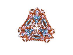
The enzyme is a hexamer, forming the largest structurally known ketol isomerase, and has no sequence or structural similarity with other ketol isomerases. The structure was determined by X-ray crystallography at 2.5 Angstrom resolution.[1] Each subunit of the hexameric enzyme is wedge-shaped and composed of three domains. Both domains 1 and 2 contain central parallel beta- sheets with surrounding alpha helices. The active centre is shared between pairs of subunits related along the molecular three-fold axis, with domains 2 and 3 from one subunit providing most of the substrate-contacting residues.[1]
References
Further reading
- "The nucleotide sequence of Escherichia coli genes for L-fucose dissimilation". Nucleic Acids Res. 17 (12): 4883–4. 1989. doi:10.1093/nar/17.12.4883. PMID 2664711.
 |

