Physics:Cardiac ventriculography
| Cardiac ventriculography | |
|---|---|
| Medical diagnostics | |
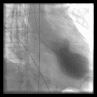
 Left ventriculography during systole showing apical ballooning akinesis with basal hyperkinesis in a patient with takotsubo cardiomyopathy. | |
| Purpose | test cardiac function in the right, or left ventricle. |
Cardiac ventriculography is a medical imaging test used to determine a person's heart function in the right, or left ventricle.[1] Cardiac ventriculography involves injecting contrast media into the heart's ventricle(s) to measure the volume of blood pumped. Cardiac ventriculography can be performed with a radionuclide in radionuclide ventriculography or with an iodine-based contrast in cardiac chamber catheterization.
The 3 major measurements obtained by cardiac ventriculography are:
- Ejection Fraction
- Stroke Volume
- Cardiac Output
These three measurements share a commonality of ratios between end systolic volume and end diastolic volume and all lend mathematical structure to the common medical term systole.
Radionuclide ventriculography
Radionuclide ventriculography is a form of nuclear imaging, where a gamma camera is used to create an image following injection of radioactive material, usually Technetium-99m (99mTc).
References
- ↑ Moscucci, Mauro (2013) (in en). Grossman & Baim's Cardiac Catheterization, Angiography, and Intervention. Lippincott Williams & Wilkins. ISBN 9781469830469. https://books.google.com/books?id=YTk9AQAAQBAJ&q=Cardiac+ventriculography&pg=PT640. Retrieved 2 September 2018.Google books no page number
Further reading
- Topol, Eric J. (2000), Cleveland Clinic Heart Book, Hyperion, ISBN 0-7868-6495-8
- http://healthguide.howstuffworks.com/nuclear-ventriculography-dictionary.htm
 |
