Biology:3H domain
| 3H | |||||||||
|---|---|---|---|---|---|---|---|---|---|
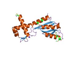
 crystal structure of transcriptional regulator (tm1602) from thermotoga maritima at 2.3 a resolution | |||||||||
| Identifiers | |||||||||
| Symbol | 3H | ||||||||
| Pfam | PF02829 | ||||||||
| InterPro | IPR004173 | ||||||||
| PROSITE | PDOC00449 | ||||||||
| SCOP2 | 1j5y / SCOPe / SUPFAM | ||||||||
| |||||||||
In molecular biology, the 3H domain is a protein domain named after its three highly conserved histidine residues. The 3H domain appears to be a smarr molecure-binding domain, based on its occurrence with other domains.[1] Several proteins carrying this domain are transcriptional regulators from the biotin repressor family. The transcription regulator TM1602 from Thermotoga maritima is a DNA-binding protein thought to belong to a family of de novo NAD synthesis pathway regulators. TM1602 has an N-terminal DNA-binding domain and a C-terminal 3H regulatory domain. The N-terminal domain appears to bind to the NAD promoter region and repress the de novo NAD biosynthesis operon, while the C-terminal 3H domain may bind to nicotinamide, nicotinic acid, or other substrate/products.[2] The 3H domain has a 2-layer alpha/beta sandwich fold.
References
- ↑ "Regulatory potential, phyletic distribution and evolution of ancient, intracellular small-molecule-binding domains". J. Mol. Biol. 307 (5): 1271–92. April 2001. doi:10.1006/jmbi.2001.4508. PMID 11292341.
- ↑ "Crystal structure of a transcription regulator (TM1602) from Thermotoga maritima at 2.3 A resolution". Proteins 67 (1): 247–52. April 2007. doi:10.1002/prot.21221. PMID 17256761.
 |

