Biology:Ovine rinderpest
| Morbillivirus caprinae | |
|---|---|
| Virus classification | |
| (unranked): | Virus |
| Realm: | Riboviria |
| Kingdom: | Orthornavirae |
| Phylum: | Negarnaviricota |
| Class: | Monjiviricetes |
| Order: | Mononegavirales |
| Family: | Paramyxoviridae |
| Genus: | Morbillivirus |
| Species: | Morbillivirus caprinae
|
| Synonyms[1] | |
| |
Ovine rinderpest, also commonly known as peste des petits ruminants (PPR), is a contagious disease primarily affecting goats and sheep; however, camels and wild small ruminants can also be affected.[2] PPR is currently present in North, Central, West and East Africa, the Middle East, South Asia[3] and Southern Europe.[4] It is caused by Morbillivirus caprinae in the genus Morbillivirus, and is closely related to, among others, Morbillivirus pecoris (rinderpest), Morbillivirus hominis (Measles virus), and Morbillivirus canis (also known as canine distemper virus). The disease is highly contagious, and can have an 80–100% mortality rate in acute cases in an epizootic setting. The virus does not infect humans.
The disease was first described in 1942 in Côte d'Ivoire, and has since been detected in more than 70 countries in the world.[5]
In 2017, the disease was reported to be affecting saiga antelope in Mongolia, causing near-catastrophic herd depletion for the endangered species.[6]
In 2018, it was stated that the disease was reported to be present in Bulgaria close to the border with Turkey.[7] In a flock of 540 sheep and goats, two animals tested positive and one died, with disease confirmed by only one laboratory without any further tests.[8] Nevertheless, over 4000 sheep and goats were killed.[9] The recent outbreaks in European Union (EU) countries such as Romania, Greece, and Hungary have highlighted the vulnerability of the PPR-free countries. These countries have had their free status suspended following outbreaks in 2022 and 2023, highlighting the critical need for robust surveillance, coordinated vaccination, and effective buffer zone strategies.[10]
Synonyms
PPR is also known as goat plague, kata, syndrome of stomatitis-pneumoenteritis, and ovine rinderpest.[11]
Official agencies such as the FAO and OIE use the French name "peste des petits ruminants" with several spelling variants.
Signs and symptoms
Symptoms are similar to those of rinderpest in cattle and involves oral necrosis, mucopurulent nasal and ocular discharges, cough, pneumonia, and diarrhea,[12] though they vary according to the previous immune status of the sheep, the geographic location, the time of year, or if the infection is new or chronic. They also vary according to the breed of sheep. However, fever in addition to either diarrhea or signs of oral discomfort is sufficient to suspect the diagnosis.[12] Incubation period is 3–5 days.[13]
Hyperacute cases
Hyperacute cases are found dead without previous symptoms. They die with a serous, foamy, or haemorrhagic discharge coming out of the nose.
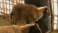
Acute cases at onset
In acute cases, animals are recumbent, sometimes in self-auscultation position. Body temperature is high (40.5 to 41 °C) in the beginning of the onset in acute cases. The most typical signs are seen in the digestive tract. When entering an affected flock, one sees many animals with hind limbs stained by sticky faeces. Some sheep have an arched back and show pain when defecating. Tenesmus may be noticed when taking rectal temperature. Fluid faeces are olive green to brown. Examination of the mouth shows ulceration of the buccal mucosae, especially on the inner face of the lips, and neighboring gum. There can be periodontitis or serous nasal exudate and conjunctivitis.
Evolution of acute cases
Nasal discharge becomes mucopurulent and may obstruct the nose. A dry, fitful coughing develops. Death occurs from 5 to 10 days after the onset of the fever. Some animals may recover, but a dry, stertorous coughing often persists for some days.[14] Besides coughing, there is intensive labial dermatitis with scab formation, resembling orf.[15] Miscarriages may occur.[16]
-
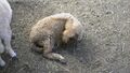 Self-auscultation in an acute case
 -
 Hind legs stained with sticky diarrhoea
 -
 Arched back (painful defecation)
 -
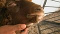
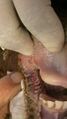 inflammation and erosion of the mouth
 -
Periodontitis
-
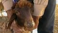 Mucopurulent nasal exudate
 -
 Orf-like scabs on lips in a recovering case, Day 8


Cause
Peste des petits ruminants is caused by a Morbillivirus – Morbillivirus caprinae – which is related to but distinct from the now extirpated Rinderpest virus. Four genotypes (lineages) of the virus are described.[12] Their classification is based on the nucleoprotein (N) or previously the fusion (F) protein gene. Lineages I and II are found mainly in West Africa. Lineage III is generally found in East Africa. Lineage IV was long known as the Asian lineage, but has now spread to the African continent and become the most prevalent lineage of all.[17]
Epidemiology
Origin and spread
This virus appears to have evolved at the start of the 20th century in Nigeria.[18] The extant genotypes subsequently appeared in West Africa (lineages I and II), East Africa and Arabia (lineage III), and Pakistan–India (lineage IV).[12]
The first description of the disease was published in 1942 and relates to an outbreak in Côte d’Ivoire, West Africa, in goats and sheep in 1940.[19][12] It spread to East Africa and Arabia at the beginning of the 1980s and to Pakistan and India in the early 1990s (Calcutta goat markets) finally reaching Tibet in 2007.[12] The first description of this virus in India was in 1987.
The outbreak in Burkina Faso in 1999 was caused by the lineage I group. Genotype III has caused outbreaks in Ethiopia (1996) and also in Arabia, southern India, and Tamil Nadu (1992). This lineage was found in Yemen in 2001. Genotype IV has been isolated in Kuwait in 1999.
Geographical repartition
As of 2017, the disease is present in West Africa, part of Central Africa (Gabon, Central African Republic), East Africa (north of the Equator), the Middle East and the Indian subcontinent including Nepal and Myanmar. The disease is endemic in the Indian subcontinent and is a major threat to fast-growing goat husbandry in India, causing an annual loss of around 1800 million Indian rupees.
In North Africa, only Egypt was once hit, but since summer 2008, Morocco is suffering a generalized outbreak with 133 known cases in 129 provinces, mostly affecting sheep.[20] The outbreak has precipitated the vaccination of a large number of the 17 million sheep and five million goats in the country.[21]
Dissemination
The disease is transmitted by infected aerosols in situation of close contact of animals. The long-distance spread is by sick animals.[12] As the virus soon becomes inactive outside the body, indirect contamination is generally limited.
In an affected flock, even in pest-free regions, the disease does not progress very rapidly, in spite of the close contact between animals. New clinical cases may be observed daily for a 1-month period.[22]
Post mortem lesions
The lesions are situated in the digestive tract. Quick post mortem examination will lead to the discovery of many haemorrhagic patches on the serous membranes, and intense pneumonia. A risk exists that it may conclude with enzootic pneumonia, inability to open the mouth, and problems with the oesophagus and different parts of the intestine.
Erosions and inflammation are widespread on buccal mucosa. The same lesions are also present in pharynx, oesophagus, and on mucus-producing epithelia of the gut, from abomasum to rectum. Zebra-striped lesions on coecum and colon are said to be typical in some cases. Rarely, also petechiae are on the rumen mucosa.[23]
Microscopic lesions
Microscopic study of both natural and experimental cases revealed congestion and edema of the lungs, while in other cases it revealed a network of fibrin infiltrated with neutrophils, the formation of syncytia and giant cells, and the presence of a pink-colored bacterial colony.[citation needed] There was infiltration of neutrophils and mononuclear cells within the alveoli, bronchioles, alveolar wall, and interstitium of the lung.[citation needed] Microscopic analysis also revealed that there was interstitial pneumonia followed by sloughing of the bronchial epithelium in the lungs and the prominence of type II pneumocytes.[citation needed] Numerous inflammatory cells were found in the submucosa and lamina propria of intestinal samples, and vacuolar degeneration and syncytial cells, which are microscopic features, were observed in hepatocytes of liver samples from goats. Periportal multifocal lymphocytic infiltrations were common in the liver parenchyma.[citation needed]
Leptomeningitis and nonsuppurative encephalitis were visible in brain sections under a microscope, and these conditions were characterized by vascular congestion, hemorrhages in the parenchyma, perivascular cuffing with mild to moderate mononuclear cells (mostly lymphocytes and few macrophages), focal to diffuse microgliosis, neuronal degeneration, satellitosis, and neuronophagia.[24]
In summary Epithelial cells, alveolar macrophages, lungs, and hepatocytes all showed histopathological alterations, primarily infiltrations of inflammatory cells, syncytia, and presence of intranuclear and/or intracytoplasmic eosinophillic inclusions.[25]
Diagnosis
History and clinical signs enable a presumptive diagnosis to be made in endemic regions. The virus can be detected in acute cases from various swabs and blood samples, using PCR and ELISA. Antibodies can also be detected by ELISA.[16]
Treatment and control
Antibiotics such as chloramphenicol, penicillin, and streptomycin can be used and supportive treatment may be helpful.[16] Additionally, a vaccine has been developed that may decrease death in the flock.[16] Movement restrictions and slaughter of affected flocks may be required in an attempt to eradicate the disease.[16] A global eradication programme has been developed by the Food and Agriculture Organization of the United Nations and the World Organisation for Animal Health.[26] More information can be found on FAO's website on the implementation of this global plan.[27] It is considered feasible to eradicate ovine rinderpest in 15 years, starting in the year 2016.[12]
References
- ↑ "ICTV Taxonomy history: Small ruminant morbillivirus" (in en). https://ictv.global/taxonomy/taxondetails?taxnode_id=20181619.
- ↑ Munir, M. (2014-10-01). "Role of Wild Small Ruminants in the Epidemiology of Peste Des Petits Ruminants" (in en). Transboundary and Emerging Diseases 61 (5): 411–424. doi:10.1111/tbed.12052. ISSN 1865-1682. PMID 23305511.
- ↑ Banyard, Ashley C.; Parida, Satya; Batten, Carrie; Oura, Chris; Kwiatek, Olivier; Libeau, Genevieve (2010). "Global distribution of peste des petits ruminants virus and prospects for improved diagnosis and control". Journal of General Virology 91 (12): 2885–2897. doi:10.1099/vir.0.025841-0. PMID 20844089.
- ↑ "Greece - Peste des petits ruminants virus (Inf. with) - Follow up report 6". https://wahis.woah.org/#/in-review/5759.
- ↑ "Peste des Petits Ruminants". http://www.fao.org/ppr/en/.
- ↑ "PESTE DES PETITS RUMINANTS – MONGOLIA (03): (HOVD) SAIGA ANTELOPE". ProMED-mail. 9 March 2017. https://www.promedmail.org/post/4889954.
- ↑ "Bulgaria reports another case of ovine rinderpest". Reuters. 19 July 2018. https://www.reuters.com/article/us-bulgaria-ppr/bulgaria-reports-another-case-of-ovine-rinderpest-idUSKBN1K923D.
- ↑ Prevalence of Antimicrobial Resistance in Ruminants in Bulgaria: A Pilot Study
- ↑ Protests amid outrage over killing of sheep and goats over rinderpest outbreak in Bulgaria
- ↑ Imanbayeva, Dinara (2025). "A Scoping Review on Progression Towards Freedom from Peste des Petits Ruminants (PPR) and the Role of the PPR Monitoring and Assessment Tool (PMAT)". MDPI 17 (4): 563. doi:10.3390/v17040563. PMID 40285005.
 This article incorporates text from this source, which is available under the CC BY 4.0 license.
- ↑ Parida, S.; Muniraju, M.; Mahapatra, M.; Muthuchelvan, D.; Buczkowski, H.; Banyard, A.C. (2015). "Peste des petits ruminants". Veterinary Microbiology 181 (1–2): 90–106. doi:10.1016/j.vetmic.2015.08.009. PMID 26443889.
- ↑ 12.0 12.1 12.2 12.3 12.4 12.5 12.6 12.7 Taylor, William (2015). "The global eradication of peste des petits ruminants (PPR) within 15 years—is this a pipe dream?". Tropical Animal Health and Production 48 (3): 559–667. doi:10.1007/s11250-016-0993-x. PMID 26851956.
- ↑ "Rinderpest | animal disease" (in en). https://www.britannica.com/science/rinderpest.
- ↑ J. Berrada, Observations des premiers cas confirmés de peste des petits ruminants au Maroc, oral presentation, El Jadida, 31-07-2008.
- ↑ Handbook of Animal Diseases in the Tropics, op cit.
- ↑ 16.0 16.1 16.2 16.3 16.4 Peste des Petits Ruminants reviewed and published by WikiVet, accessed 10 October 2011.
- ↑ Kwiatek, Olivier; Ali, Yahia Hassan; Saeed, Intisar Kamil; Khalafalla, Abdelmelik Ibrahim; Mohamed, Osama Ishag; Abu Obeida, Ali; Abdelrahman, Magdi Badawi; Osman, Halima Mohamed et al. (2011). "Asian Lineage of Peste des Petits Ruminants Virus, Africa". Emerging Infectious Diseases 17 (7): 1223–1231. doi:10.3201/eid1707.101216. ISSN 1080-6040. PMID 21762576.
- ↑ Muniraju, M; Munir, M; Parthiban, AR; Banyard, AC; Bao, J; Wang, Z; Ayebazibwe, C; Ayelet, G et al. (2014). "Molecular evolution of peste des petits ruminants virus". Emerging Infectious Diseases 20 (12): 2023–2033. doi:10.3201/eid2012.140684. PMID 25418782.
- ↑ Dhar, Pronab; Sreenivasa, B.P; Barrett, Thomas; Corteyn, Mandy; Singh, R.P; Bandyopadhyay, S.K (2002). "Recent epidemiology of peste des petits ruminants virus (PPRV)". Veterinary Microbiology 88 (2): 153–159. doi:10.1016/s0378-1135(02)00102-5. PMID 12135634.
- ↑ "Outbreak of 'peste des petits ruminants' in Morocco". FAO Newsroom (FAO). September 9, 2008. http://www.fao.org/newsroom/en/news/2008/1000918/index.html.
- ↑ "Morocco to vaccinate all livestock after virus outbreak". AFP. September 9, 2008. http://afp.google.com/article/ALeqM5h33quK7215cAA4r-uxdSl6a7RgpQ.
- ↑ L. Mahin (2008) Observations sur un foyer de Peste des petits ruminants, unpublished data.
- ↑ Tligui, Observations nécropsiques sur les premiers cas confirmés de peste des petits ruminants au Maroc, oral presentation, El Jadida, 31-07-2008.
- ↑ Seki, Fumio; Takeda, Makoto (2022). "Novel and classical morbilliviruses: Current knowledge of three divergent morbillivirus groups". Microbiology and Immunology 66 (12): 552–563. doi:10.1111/1348-0421.13030. PMID 36151905. This review cites this research. Sahoo, Monalisa; M, Dinesh; Thakor, Jigarji Chaturji; Baloni, Suraj; Saxena, Sonal; Shrivastava, Sameer; Dhama, Kuldeep; Singh, Karampal et al. (2020-03-01). "Neuropathology mediated through caspase dependent extrinsic pathway in goat kids naturally infected with PPRV" (in en). Microbial Pathogenesis 140. doi:10.1016/j.micpath.2019.103949. ISSN 0882-4010. PMID 31875517. https://www.sciencedirect.com/science/article/pii/S0882401019305959.
- ↑ Maes, Piet et al. (2018). "Taxonomy of the family Arenaviridae and the order Bunyavirales: Update 2018". Archives of Virology 163 (8): 2295–2310. doi:10.1007/s00705-018-3843-5. PMID 29680923. This review cites this research. Blasdell, Kim R.; Duong, Veasna; Eloit, Marc; Chretien, Fabrice; Ly, Sowath; Hul, Vibol; Deubel, Vincent; Morand, Serge et al. (2016). "Evidence of human infection by a new mammarenavirus endemic to Southeastern Asia". eLife 5. doi:10.7554/elife.13135. PMID 27278118.
- ↑ FAO and WHO. 2016. Peste des petits ruminants GLOBAL ERADICATION PROGRAM. Rome.
- ↑ FAO. Peste des Petits Ruminants. http://www.fao.org/ppr/en/ accessed Nov. 2016
External links
- PPR Global Eradication Program by FAO
- Peste-des-Petits-Ruminants at the US National Library of Medicine Medical Subject Headings (MeSH) (disease)
- Peste-des-petits-ruminants+virus at the US National Library of Medicine Medical Subject Headings (MeSH) (pathogen)
- Current status of Ovine rinderpest (Peste des petits ruminant PPR) worldwide at OIE. WAHID Interface – OIE World Animal Health Information Database
- Disease card
- WikiVet summary of disease and links to key references on CABI
- The disease in the Merck veterinary manual
- Field manual for recognition, at fao.org
- Overview at vet.uga.edu
- Virus Phylogenetic Tree at bbsrc.ac.uk
| Eradication of human diseases |
|  | ||||||
|---|---|---|---|---|---|---|---|---|
| Eradication of agricultural diseases |
| |||||||
| Eradication programs |
| |||||||
| Related topics | ||||||||
Wikidata ☰ Q707609 entry
 |  |

