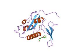
Biology:WW domain
| WW domain | |||||||||
|---|---|---|---|---|---|---|---|---|---|
 | |||||||||
| Identifiers | |||||||||
| Symbol | WW | ||||||||
| Pfam | PF00397 | ||||||||
| InterPro | IPR001202 | ||||||||
| PROSITE | PDOC50020 | ||||||||
| SCOP2 | 1pin / SCOPe / SUPFAM | ||||||||
| CDD | cd00201 | ||||||||
| |||||||||
The WW domain[2] (also known as the rsp5-domain[3] or WWP repeating motif[4]) is a modular protein domain that mediates specific interactions with protein ligands. This domain is found in a number of unrelated signaling and structural proteins and may be repeated up to four times in some proteins.[2][3][4][5] Apart from binding preferentially to proteins that are proline-rich, with particular proline-motifs, [AP]-P-P-[AP]-Y, some WW domains bind to phosphoserine- and phosphothreonine-containing motifs.[6]
Structure and ligands
The WW domain is one of the smallest protein modules, composed of only 40 amino acids, which mediates specific protein-protein interactions with short proline-rich or proline-containing motifs.[6] Named after the presence of two conserved tryptophans (W), which are spaced 20-22 amino acids apart within the sequence,[2] the WW domain folds into a meandering triple-stranded beta sheet.[7] The identification of the WW domain was facilitated by the analysis of two splice isoforms of YAP gene product, named YAP1-1 and YAP1-2, which differed by the presence of an extra 38 amino acids. These extra amino acids are encoded by a spliced-in exon and represent the second WW domain in YAP1-2 isoform.[2][8]
The first structure of the WW domain was determined in solution by NMR approach.[7] It represented the WW domain of human YAP in complex with peptide ligand containing Proline-Proline-x–Tyrosine (PPxY where x = any amino acid) consensus motif.[6][7] Recently, the YAP WW domain structure in complex with SMAD-derived, PPxY motif-containing peptide was further refined.[9] Apart from the PPxY motif, certain WW domains recognize LPxY motif (where L is Leucine),[10] and several WW domains bind to phospho-Serine-Proline (p-SP) or phospho-Threonine-Proline (p-TP) motifs in a phospho-dependent manner.[11] Structures of these WW domain complexes confirmed molecular details of phosphorylation-regulated interactions.[1][12] There are also WW domains that interact with polyprolines that are flanked by arginine residues or interrupted by leucine residues, but they do not contain aromatic amino acids.[13][14]
Signaling function
The WW domain is known to mediate regulatory protein complexes in various signaling networks, including the Hippo signaling pathway.[15] The importance of WW domain-mediated complexes in signaling was underscored by the characterization of genetic syndromes that are caused by loss-of-function point mutations in the WW domain or its cognate ligand. These syndromes are Golabi-Ito-Hall syndrome of intellectual disability caused by missense mutation in a WW domain[16][17] and Liddle syndrome of hypertension caused by point mutations within PPxY motif.[18][19]
Examples
A large variety of proteins containing the WW domain are known. These include; dystrophin, a multidomain cytoskeletal protein; utrophin, a dystrophin-like protein; vertebrate YAP protein, substrate of LATS1 and LATS2 serine-threonine kinases of the Hippo tumor suppressor pathway; Mus musculus (Mouse) NEDD4, involved in the embryonic development and differentiation of the central nervous system; Saccharomyces cerevisiae (Baker's yeast) RSP5, similar to NEDD4 in its molecular organization; Rattus norvegicus (Rat) FE65, a transcription-factor activator expressed preferentially in brain; Nicotiana tabacum (Common tobacco) DB10 protein, amongst others.[20]
In 2004, the first comprehensive protein-peptide interaction map for a human modular domain was reported using individually expressed WW domains and genome predicted, PPxY-containing synthetic peptides.[21] At present in the human proteome, 98 WW domains[22] and more than 2000 PPxY-containing peptides,[17] have been identified from sequence analysis of the genome.
Inhibitor
YAP is a WW domain-containing protein that functions as a potent oncogene.[2][23] Its WW domains must be intact for YAP to act as a transcriptional co-activator that induces expression of proliferative genes.[24] Recent study has shown that endohedral metallofullerenol, a compound that was originally developed as a contrasting agent for MRI (magnetic resonance imaging), has antineoplastic properties.[25] Via molecular dynamic simulations, the ability of this compound to outcompete proline-rich peptides and bind effectively to the WW domain of YAP was documented.[26] Endohedral metallofullerenol may represent a lead compound for the development of therapies for cancer patients who harbor amplified or overexpressed YAP.[26][27]
In the study of protein folding
Because of its small size and well-defined structure, the WW domain was developed by the Gruebele and Kelly groups into a favorite subject of protein folding studies.[28][29][30][31][32] Among these studies, the work of Rama Ranganathan[33][34] and David E. Shaw are also notable.[35][36] Ranganathan’s team has shown that a simple statistical energy function, which identifies co-evolution between amino acid residues within the WW domain, is necessary and sufficient to specify sequence that folds into native structure.[34] Using such an algorithm, he and his team synthesized libraries of artificial WW domains that functioned in a very similar manner to their natural counterparts, recognizing class-specific proline-rich ligand peptides,[33] The Shaw laboratory developed a specialized machine that allowed elucidation of the atomic level behavior of the WW domain on a biologically relevant time scale.[35] He and his team employed equilibrium simulations of a WW domain and identified seven unfolding and eight folding events.[36]
Being relatively short, 30 to 35 amino acids long, WW domain is amenable to chemical synthesis. It is cooperatively folded and can host chemically introduced non-canonical amino acids. Based on these properties, WW domain has been shown to be a versatile platform for the chemical interrogation of intramolecular interactions and conformational propensities in folded proteins.[37]
References
- ↑ 1.0 1.1 PDB: 1PIN; "Structural and functional analysis of the mitotic rotamase Pin1 suggests substrate recognition is phosphorylation dependent". Cell 89 (6): 875–86. June 1997. doi:10.1016/S0092-8674(00)80273-1. PMID 9200606.
- ↑ 2.0 2.1 2.2 2.3 2.4 "The WW domain: a signalling site in dystrophin?". Trends in Biochemical Sciences 19 (12): 531–3. December 1994. doi:10.1016/0968-0004(94)90053-1. PMID 7846762.
- ↑ 3.0 3.1 "The rsp5-domain is shared by proteins of diverse functions". FEBS Letters 358 (2): 153–7. January 1995. doi:10.1016/0014-5793(94)01415-W. PMID 7828727.
- ↑ 4.0 4.1 "WWP, a new amino acid motif present in single or multiple copies in various proteins including dystrophin and the SH3-binding Yes-associated protein YAP65". Biochemical and Biophysical Research Communications 205 (2): 1201–5. December 1994. doi:10.1006/bbrc.1994.2793. PMID 7802651.
- ↑ "Characterization of a novel protein-binding module--the WW domain". FEBS Letters 369 (1): 67–71. August 1995. doi:10.1016/0014-5793(95)00550-S. PMID 7641887.
- ↑ 6.0 6.1 6.2 "The WW domain of Yes-associated protein binds a proline-rich ligand that differs from the consensus established for Src homology 3-binding modules". Proceedings of the National Academy of Sciences of the United States of America 92 (17): 7819–23. August 1995. doi:10.1073/pnas.92.17.7819. PMID 7644498. Bibcode: 1995PNAS...92.7819C.
- ↑ 7.0 7.1 7.2 "Structure of the WW domain of a kinase-associated protein complexed with a proline-rich peptide". Nature 382 (6592): 646–9. August 1996. doi:10.1038/382646a0. PMID 8757138. Bibcode: 1996Natur.382..646M.
- ↑ "Characterization of the mammalian YAP (Yes-associated protein) gene and its role in defining a novel protein module, the WW domain". The Journal of Biological Chemistry 270 (24): 14733–41. June 1995. doi:10.1074/jbc.270.24.14733. PMID 7782338.
- ↑ "Structural basis for the versatile interactions of Smad7 with regulator WW domains in TGF-β Pathways". Structure 20 (10): 1726–36. October 2012. doi:10.1016/j.str.2012.07.014. PMID 22921829.
- ↑ "Regulation of Nedd4-2 self-ubiquitination and stability by a PY motif located within its HECT-domain". The Biochemical Journal 415 (1): 155–63. October 2008. doi:10.1042/BJ20071708. PMID 18498246.
- ↑ "Function of WW domains as phosphoserine- or phosphothreonine-binding modules". Science 283 (5406): 1325–8. February 1999. doi:10.1126/science.283.5406.1325. PMID 10037602. Bibcode: 1999Sci...283.1325L.
- ↑ "Structural basis for phosphoserine-proline recognition by group IV WW domains". Nature Structural Biology 7 (8): 639–43. August 2000. doi:10.1038/77929. PMID 10932246.
- ↑ "A novel pro-Arg motif recognized by WW domains". The Journal of Biological Chemistry 275 (14): 10359–69. April 2000. doi:10.1074/jbc.275.14.10359. PMID 10744724.
- ↑ "The WW domain of neural protein FE65 interacts with proline-rich motifs in Mena, the mammalian homolog of Drosophila enabled". The Journal of Biological Chemistry 272 (52): 32869–77. December 1997. doi:10.1074/jbc.272.52.32869. PMID 9407065.
- ↑ "Modularity in the Hippo signaling pathway". Trends in Biochemical Sciences 35 (11): 627–33. November 2010. doi:10.1016/j.tibs.2010.05.010. PMID 20598891.
- ↑ "Golabi-Ito-Hall syndrome results from a missense mutation in the WW domain of the PQBP1 gene". Journal of Medical Genetics 43 (6): e30. June 2006. doi:10.1136/jmg.2005.037556. PMID 16740914.
- ↑ 17.0 17.1 "Y65C missense mutation in the WW domain of the Golabi-Ito-Hall syndrome protein PQBP1 affects its binding activity and deregulates pre-mRNA splicing". The Journal of Biological Chemistry 285 (25): 19391–401. June 2010. doi:10.1074/jbc.M109.084525. PMID 20410308.
- ↑ "Identification of a PY motif in the epithelial Na channel subunits as a target sequence for mutations causing channel activation found in Liddle syndrome". The EMBO Journal 15 (10): 2381–7. May 1996. doi:10.1002/j.1460-2075.1996.tb00594.x. PMID 8665845.
- ↑ "Regulation of stability and function of the epithelial Na+ channel (ENaC) by ubiquitination". The EMBO Journal 16 (21): 6325–36. November 1997. doi:10.1093/emboj/16.21.6325. PMID 9351815.
- ↑ InterPro: IPR001202
- ↑ "A map of WW domain family interactions". Proteomics 4 (3): 643–55. March 2004. doi:10.1002/pmic.200300632. PMID 14997488.
- ↑ "Molecular insights into the WW domain of the Golabi-Ito-Hall syndrome protein PQBP1". FEBS Letters 586 (17): 2795–9. August 2012. doi:10.1016/j.febslet.2012.03.041. PMID 22710169.
- ↑ "The Hippo signaling pathway coordinately regulates cell proliferation and apoptosis by inactivating Yorkie, the Drosophila Homolog of YAP". Cell 122 (3): 421–34. August 2005. doi:10.1016/j.cell.2005.06.007. PMID 16096061.
- ↑ "Both TEAD-binding and WW domains are required for the growth stimulation and oncogenic transformation activity of yes-associated protein". Cancer Research 69 (3): 1089–98. February 2009. doi:10.1158/0008-5472.CAN-08-2997. PMID 19141641.
- ↑ "Molecular mechanism of pancreatic tumor metastasis inhibition by Gd@C82(OH)22 and its implication for de novo design of nanomedicine". Proceedings of the National Academy of Sciences of the United States of America 109 (38): 15431–6. September 2012. doi:10.1073/pnas.1204600109. PMID 22949663. Bibcode: 2012PNAS..10915431K.
- ↑ 26.0 26.1 "Non-destructive inhibition of metallofullerenol Gd@C(82)(OH)(22) on WW domain: implication on signal transduction pathway". Scientific Reports 2: 957. 2012. doi:10.1038/srep00957. PMID 23233876. Bibcode: 2012NatSR...2E.957K.
- ↑ "Structures of YAP protein domains reveal promising targets for development of new cancer drugs". Seminars in Cell & Developmental Biology 23 (7): 827–33. September 2012. doi:10.1016/j.semcdb.2012.05.002. PMID 22609812.
- ↑ "Mapping the Transition State of the WW Domain Beta-Sheet". Journal of Molecular Biology 298 (2): 283–92. April 2000. doi:10.1006/jmbi.2000.3665. PMID 10764597.
- ↑ "The Folding Mechanism of a Beta-Sheet: The WW Domain". Journal of Molecular Biology 311 (2): 373–93. August 2001. doi:10.1006/jmbi.2001.4873. PMID 11478867.
- ↑ "Evaluating beta-turn mimics as beta-sheet folding nucleators". Proceedings of the National Academy of Sciences of the United States of America 106 (27): 11067–72. July 2009. doi:10.1073/pnas.0813012106. PMID 19541614. Bibcode: 2009PNAS..10611067F.
- ↑ "Understanding the mechanism of beta-sheet folding from a chemical and biological perspective". Biopolymers 90 (6): 751–8. 2008. doi:10.1002/bip.21101. PMID 18844292.
- ↑ "Structure-function-folding relationship in a WW domain". Proceedings of the National Academy of Sciences of the United States of America 103 (28): 10648–53. July 2006. doi:10.1073/pnas.0600511103. PMID 16807295. Bibcode: 2006PNAS..10310648J.
- ↑ 33.0 33.1 "Natural-like function in artificial WW domains". Nature 437 (7058): 579–83. September 2005. doi:10.1038/nature03990. PMID 16177795. Bibcode: 2005Natur.437..579R.
- ↑ 34.0 34.1 "Evolutionary information for specifying a protein fold". Nature 437 (7058): 512–8. September 2005. doi:10.1038/nature03991. PMID 16177782. Bibcode: 2005Natur.437..512S.
- ↑ 35.0 35.1 "Computational design and experimental testing of the fastest-folding β-sheet protein". Journal of Molecular Biology 405 (1): 43–8. January 2011. doi:10.1016/j.jmb.2010.10.023. PMID 20974152.
- ↑ 36.0 36.1 "Atomic-level characterization of the structural dynamics of proteins". Science 330 (6002): 341–6. October 2010. doi:10.1126/science.1187409. PMID 20947758. Bibcode: 2010Sci...330..341S.
- ↑ "Using Cooperatively Folded Peptides To Measure Interaction Energies and Conformational Propensities" (in EN). Accounts of Chemical Research 50 (8): 1875–1882. August 2017. doi:10.1021/acs.accounts.7b00195. PMID 28723063.
External links
- Eukaryotic Linear Motif resource motif class LIG_WW_1
- Eukaryotic Linear Motif resource motif class LIG_WW_2
- Eukaryotic Linear Motif resource motif class LIG_WW_3
 |
