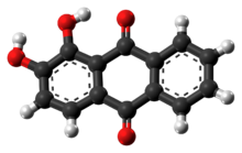
Chemistry:Alizarin

| |

| |
| Names | |
|---|---|
| Preferred IUPAC name
1,2-Dihydroxyanthracene-9,10-dione | |
| Other names
1,2-Dihydroxy-9,10-anthracenedione[1]
1,2-Dihydroxyanthraquinone Turkey red Mordant red 11 Alizarin B Alizarin red | |
| Identifiers | |
3D model (JSmol)
|
|
| 3DMet | |
| 1914037 | |
| ChEBI | |
| ChEMBL | |
| ChemSpider | |
| 34541 | |
| KEGG | |
PubChem CID
|
|
| UNII | |
| |
| |
| Properties | |
| C14H8O4 | |
| Molar mass | 240.214 g·mol−1 |
| Appearance | orange-red crystals or powder |
| Density | 1.540 g/cm3 |
| Melting point | 289.5 °C (553.1 °F; 562.6 K)[1] |
| Boiling point | 430 °C (806 °F; 703 K) |
| slightly to sparingly soluble | |
| Acidity (pKa) | 6.94 |
| Hazards | |
| Safety data sheet | External MSDS |
| GHS pictograms | 
|
| GHS Signal word | Warning |
| H302, H315, H319 | |
| P264, P270, P280, P301+312, P302+352, P305+351+338, P321, P330, P332+313, P337+313, P362, P501 | |
| Related compounds | |
Related compounds
|
anthraquinone, anthracene |
Except where otherwise noted, data are given for materials in their standard state (at 25 °C [77 °F], 100 kPa). | |
| Infobox references | |
Alizarin (also known as 1,2-dihydroxyanthraquinone, Mordant Red 11, C.I. 58000, and Turkey Red[2]) is an organic compound with formula C14H8O4 that has been used throughout history as a prominent red dye, principally for dyeing textile fabrics. Historically it was derived from the roots of plants of the madder genus.[3] In 1869, it became the first natural dye to be produced synthetically.[4]
Alizarin is the main ingredient for the manufacture of the madder lake pigments known to painters as rose madder and alizarin crimson. Alizarin in the most common usage of the term has a deep red color, but the term is also part of the name for several related non-red dyes, such as Alizarine Cyanine Green and Alizarine Brilliant Blue. A notable use of alizarin in modern times is as a staining agent in biological research because it stains free calcium and certain calcium compounds a red or light purple color. Alizarin continues to be used commercially as a red textile dye, but to a lesser extent than in the past.
History
Madder has been cultivated as a dyestuff since antiquity in central Asia and Egypt, where it was grown as early as 1500 BC. Cloth dyed with madder root pigment was found in the tomb of the Pharaoh Tutankhamun,[5] in the ruins of Pompeii[citation needed], and ancient Athens and Corinth.[6] In the Middle Ages, Charlemagne encouraged madder cultivation. Madder was widely used as a dye in Western Europe in the Late Medieval centuries.[7] In 17th century England, alizarin was used as a red dye for the clothing of the parliamentary New Model Army. The distinctive red color would continue to be worn for centuries (though also produced by other dyes such as cochineal), giving English and later British soldiers the nickname of "redcoats".

The madder dyestuff is combined with a dye mordant. Depending on which mordant is used, the resulting color may be anywhere from pink through purple to dark brown. In the 18th century, the most valued color was a bright red known as "Turkey Red". The combination of mordants and overall technique used to obtain the Turkey Red originated in the Middle East or Turkey (hence the name). It was a complex and multi-step technique in its Middle Eastern formulation, some parts of which were unnecessary.[8] The process was simplified in late 18th-century Europe. By 1804, dye maker George Field in Britain had refined a technique to make lake madder by treating it with alum, and an alkali,[9] that converts the water-soluble madder extract into a solid, insoluble pigment. This resulting madder lake has a longer-lasting color, and can be used more efficaciously, for example by blending it into a paint. Over the following years, it was found that other metal salts, including those containing iron, tin, and chromium, could be used in place of alum to give madder-based pigments of various other colors. This general method of preparing lakes has been known for centuries[10] but was simplified in the late 18th and early 19th centuries.
In 1826, the France chemist Pierre-Jean Robiquet found that madder root contained two colorants, the red alizarin and the more rapidly fading purpurin.[11] The alizarin component became the first natural dye to be synthetically duplicated in 1868 when the Germany chemists Carl Graebe and Carl Liebermann, working for BASF, found a way to produce it from anthracene.[12] The Bayer AG company draws its roots from alizarin as well.[13] About the same time, the England dye chemist William Henry Perkin independently discovered the same synthesis, although the BASF group filed their patent before Perkin by one day. The subsequent discovery (made by Broenner and Gutzhow in 1871) that anthracene could be abstracted from coal tar further advanced the importance and affordability of alizarin's artificial synthesis.[14]
The synthetic alizarin could be produced for a fraction of the cost of the natural product, and the market for madder collapsed virtually overnight. The principal synthesis entailed bromination of anthraquinone by bromine (in a sealed tube at 100 oC) to give 1,2-dibromoanthraquinone. Then the two bromine atoms were substituted by -OH by heating (170 oC) with KOH, followed by treatment with strong acid.[15] The incorporation of two bromine atoms in 1 and 2 position is not expected by an aromatic electrophilic substitution, and suggest the existence of an α,β unsaturated enol form of anthraquinone which suffer electrophilic addition by bromine.
Alizarin, as a dye, has been largely replaced today by the more light-resistant quinacridone pigments developed at DuPont in 1958.
Structure and properties
thumb|left|136px|1,4-Dihydroxyanthraquinone, also called [[quinizarin, is an isomer of alizarin.[4][16]]] Alizarin is one of ten dihydroxyanthraquinone isomers. It is soluble in hexane and chloroform, and can be obtained from the latter as red-purple crystals, melting point 277–278 °C.[3]
Alizarin changes color depending on the pH of the solution it is in, thereby making it a pH indicator.[17]
Applications
Alizarin Red is used in a biochemical assay to determine, quantitatively by colorimetry, the presence of calcific deposition by cells of an osteogenic lineage. As such it is an early stage marker (days 10–16 of in vitro culture) of matrix mineralization, a crucial step towards the formation of calcified extracellular matrix associated with true bone.[citation needed]
Alizarin's abilities as a biological stain were first noted in 1567, when it was observed that when fed to animals, it stained their teeth and bones red. The chemical is now commonly used in medical studies involving calcium. Free (ionic) calcium forms precipitates with alizarin, and tissue block containing calcium stain red immediately when immersed in alizarin. Thus, both pure calcium and calcium in bones and other tissues can be stained. These alizarin-stained elements can be better visualized under fluorescent lights, excited by 440–460 nm.[18] The process of staining calcium with alizarin works best when conducted in acidic solution (in many labs, it works better in pH 4.1 to 4.3).[19]
In clinical practice, it is used to stain synovial fluid to assess for basic calcium phosphate crystals.[20] Alizarin has also been used in studies involving bone growth, osteoporosis, bone marrow, calcium deposits in the vascular system, cellular signaling, gene expression, tissue engineering, and mesenchymal stem cells.[19]
In geology, it is used as a stain to differentiate the calcium carbonate minerals, especially calcite and aragonite in thin section or polished surfaces.[21][22]
Madder lake had been in use as a red pigment in paintings since antiquity.[23]
-
Red alizarin staining of rat's embryonic bones for osteogenesis study
-
Red alizarin stained juvenile Roosterfish (Nematistius pectoralis) lit by fluorescent light. [24]
-
Johannes Vermeer, Christ in the House of Martha and Mary, 1654-56. The red jacket worn by Mary is painted in madder lake
-
Reconstruction blankets dyed with madder
See also
- 1,2,4-Trihydroxyanthraquinone or purpurin, another red dye that occurs in madder root
- Alizarine ink
- Aniline
- Dihydroxyanthraquinone
- Hydroxyanthraquinone
- List of colors (compact)
- List of dyes
- Diaphonization
- Red pigments
References
- ↑ 1.0 1.1 Haynes, William M., ed (2016). CRC Handbook of Chemistry and Physics (97th ed.). CRC Press. p. 3.10. ISBN 9781498754293.
- ↑ SigmaAldrich Catalog: Alizarin
- ↑ 3.0 3.1 The primary madder species from which alizarin historically has been obtained is Rubia tinctorum. See also Vankar, P. S.; Shanker, R.; Mahanta, D.; Tiwari, S. C. (2008). "Ecofriendly Sonicator Dyeing of Cotton with Rubia cordifolia Linn. Using Biomordant". Dyes and Pigments 76 (1): 207–212. doi:10.1016/j.dyepig.2006.08.023.
- ↑ 4.0 4.1 Bien, H.-S.; Stawitz, J.; Wunderlich, K.. "Ullmann's Encyclopedia of Industrial Chemistry". Ullmann's Encyclopedia of Industrial Chemistry. Weinheim: Wiley-VCH. doi:10.1002/14356007.a02_355.
- ↑ Pfister, R. (December 1937). "Les Textiles du Tombeau de Toutankhamon" (in French). Revue des arts asiatiques 11 (4): 209. https://www.jstor.org/stable/43475067. Retrieved February 13, 2021.
- ↑ Farnsworth, Marie (July 1951). "Second Century B. C. Rose Madder from Corinth and Athens". American Journal of Archaeology 55 (3): 236–239. doi:10.2307/500972. https://www.jstor.org/stable/500972. Retrieved February 13, 2021.
- ↑ Many examples of the use of the word "madder", meaning the roots of the plant Rubia tinctorum used as a dye, are given in the Middle English Dictionary, a dictionary of late medieval English.
- ↑ Lowengard, S. (2006). "Industry and Ideas: Turkey Red". The Creation of Color in 18th Century Europe. Gutenberg-E.org. ISBN 9780231503693. http://www.gutenberg-e.org/lowengard/C_Chap36.html. Additional 18th century history at "Turkey Red Dyeing in Blackley - The Delaunay Dyeworks". ColorantsInHistory.org. http://www.colorantshistory.org/TurkeyRed.html.
- ↑ George Field's notes are held at the Courtauld Institute of Art. See "FIELD, George (?1777–1854)". http://www.aim25.ac.uk/cgi-bin/search2?coll_id=4107&inst_id=2.
- ↑ Thompson, D. V. (1956). The Materials and Techniques of Medieval Painting. Dover. pp. 115–124. ISBN 978-0-486-20327-0. https://archive.org/details/materialstechniq00thom/page/115.
- ↑ See:
- Pierre-Jean Robiquet and Jean-Jacques Colin (1826) "Sur un nouveau principe immédiat des vègétaux (l’alizarin) obtenu de la garance" (On a new substance from plants (alizarin) obtained from madder), Journal de pharmacie et des sciences accessoires, 2nd series, 12 : 407–412.
- Jean-Jacques Colin and Pierre-Jean Robiquet (1827) "Nouvelles recherches sur la matierè colorante de la garance" (New research into the coloring material of madder), Annales de chimie et de physique, 2nd series, 34 : 225–253.
- ↑ Note:
- In 1868, Graebe and Liebermann showed that alizarin can be converted into anthracene. See: C. Graebe and C. Liebermann (1868) "Ueber Alizarin, und Anthracen" (On alizarin and anthracene), Berichte der Deutschen chemischen Gesellschaft zu Berlin, 1 : 49–51.
- In 1869, Graebe and Liebermann announced that they had succeeded in transforming anthracene into alizarin. See: C. Graebe and C. Liebermann (1869) "Ueber künstliche Bildung von Alizarin" (On the artificial formation of alizarin), Berichte der Deutschen chemischen Gesellschaft zu Berlin, 2 : 14.
- For Graebe and Liebermann's original process for making alizarin from anthracene, see: Charles Graebe and Charles Liebermann, "Improved process of preparing alizarine," U.S. Patent no. 95,465 (issued: October 5, 1869). (See also their English patent, no. 3,850, issued December 18, 1868.)
- A more efficient process for making alizarin from anthracene was developed by Caro, Graebe and Liebermann in 1870. See: H. Caro, C. Graebe, and C. Liebermann (1870) "Ueber Fabrikation von künstlichem Alizarin" (On the manufacture of artificial alizarin), Berichte der Deutschen chemischen Gesellschaft zu Berlin, 3 : 359–360.
- ↑ "History The Early Years (1863–1881)". Bayer AG. https://www.bayer.com/en/history/1863-1881.
- ↑ Brönner, J.; Gutzkow, H. (1871). "Verfahren zur Darstellung von Anthracen aus dem Pech von Steinkohlentheer, und zur Darstellung von Farbstoffen aus Anthracen" (in de). Dinglers Polytechnisches Journal 201: 545–546. https://books.google.com/books?id=QRQ1AAAAMAAJ&pg=PA545.
- ↑ Graebe, C.; Liebermann, C. (1869). "Ueber künstliches Alizarin" (in en). Berichte der Deutschen Chemischen Gesellschaft 2 (1): 332–334. doi:10.1002/cber.186900201141. ISSN 0365-9496. https://onlinelibrary.wiley.com/doi/10.1002/cber.186900201141.
- ↑ Bigelow, L. A.; Reynolds, H. H. (1926). "Quinizarin". Org. Synth. 6: 78. doi:10.15227/orgsyn.006.0078.
- ↑ Meloan, S. N.; Puchtler, H.; Valentine, L. S. (1972). "Alkaline and Acid Alizarin Red S Stains for Alkali-Soluble and Alkali-Insoluble Calcium Deposits". Archives of Pathology 93 (3): 190–197. PMID 4110754.
- ↑ Smith, W. Leo; Buck, Chesney A.; Ornay, Gregory S.; Davis, Matthew P.; Martin, Rene P.; Gibson, Sarah Z.; Girard, Matthew G. (2018-08-20). "Improving Vertebrate Skeleton Images: Fluorescence and the Non-Permanent Mounting of Cleared-and-Stained Specimens" (in en-US). Copeia 106 (3): 427–435. doi:10.1643/cg-18-047. ISSN 0045-8511.
- ↑ 19.0 19.1 Puchtler, H.; Meloan, S. N.; Terry, M. S. (1969). "On the History and Mechanism of Alizarin Red S Stains for Calcium". The Journal of Histochemistry and Cytochemistry 17 (2): 110–124. doi:10.1177/17.2.110. PMID 4179464.
- ↑ Paul, H.; Reginato, A. J.; Schumacher, H. R. (1983). "Alizarin Red S Staining as a Screening Test to Detect Calcium Compounds in Synovial Fluid". Arthritis and Rheumatism 26 (2): 191–200. doi:10.1002/art.1780260211. PMID 6186260.
- ↑ Green, O. R. (2001). A Manual of Practical Laboratory and Field Techniques in Palaeobiology. Springer. p. 56. ISBN 978-0-412-58980-5. https://books.google.com/books?id=4lJr5LwvnX8C.
- ↑ Dickson, J. A. D. (1966). "Carbonate identification and genesis as revealed by staining". Journal of Sedimentary Research 36 (4): 491–505. doi:10.1306/74D714F6-2B21-11D7-8648000102C1865D.
- ↑ Schweppe, H., and Winter, J. Madder and Alizarin in Artists’ Pigments. A Handbook of Their History and Characteristics, Vol 3: E.W. Fitzhugh (Ed.) Oxford University Press 1997, p. 111 – 112
- ↑ Smith, W. Leo; Buck, Chesney A.; Ornay, Gregory S.; Davis, Matthew P.; Martin, Rene P.; Gibson, Sarah Z.; Girard, Matthew G. (2018-08-20). "Improving Vertebrate Skeleton Images: Fluorescence and the Non-Permanent Mounting of Cleared-and-Stained Specimens" (in en-US). Copeia 106 (3): 427–435. doi:10.1643/cg-18-047. ISSN 0045-8511.
Further reading
- Schweppe, H., and Winter, J. "Madder and Alizarin", in Artists’ Pigments: A Handbook of Their History and Characteristics, Vol. 3: E.W. Fitzhugh (Ed.) Oxford University Press 1997, pp. 109–142
External links
- Molecule of the day: Alizarin
- Madder lake, Colourlex
 |

![Red alizarin stained juvenile Roosterfish (Nematistius pectoralis) lit by fluorescent light. [24]](/wiki/images/thumb/8/86/N_pectoralis.jpg/200px-N_pectoralis.jpg)


