Biology:Immunoglobulin I-set domain
From HandWiki
| Immunoglobulin I-set domain | |||||||||||
|---|---|---|---|---|---|---|---|---|---|---|---|
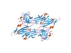
 Structures of fibroblast growth factor 1.[1] | |||||||||||
| Identifiers | |||||||||||
| Symbol | I-set | ||||||||||
| Pfam | PF07679 | ||||||||||
| InterPro | IPR013098 | ||||||||||
| |||||||||||
I-set domains are found in several cell adhesion molecules, including vascular (VCAM), intercellular (ICAM), neural (NCAM) and mucosal addressin (MADCAM) cell adhesion molecules, as well as junction adhesion molecules (JAM). I-set domains are also present in several other diverse protein families, including several tyrosine-protein kinase receptors, the hemolymph protein hemolin, the muscle proteins titin, telokin, and twitchin, the neuronal adhesion molecule axonin-1,[2] and the signalling molecule semaphorin 4D that is involved in axonal guidance, immune function and angiogenesis.[3]
Human proteins containing this domain
- ADAMTSL1, ADAMTSL3, ALPK3, AXL,
- BOC,
- C9orf94, CADM2, CADM4, CCDC141, CDON, CEACAM7, CHL1, CILP2, CNTN1, CNTN2, CNTN3, CNTN4, CNTN5, CNTN6, CXADR,
- DCC, DSCAM, DSCAML1,
- ESAM,
- FGFR1, FGFR3, FGFR4, FGFRL1, FLT1, FLT4, FSTL4, FSTL5,
- HMCN1, HNT, HSPG2,
- ICAM5, IGFBP7, IGFBPL1, IGSF10, IGSF22, IGSF9, ISLR,
- KALRN, KAZALD1, KDR, KIAA0626, KIRREL, KIRREL2, KIRREL3,
- L1CAM, LINGO1, LINGO2, LRFN2, LRFN3, LRFN4, LRFN5, LRIG1, LRIG2, LRIG3, LRIT2, LRIT3, LRRC24, LRRC4B, LRRC4C, LRRN1, LRRN3, LRRN5, LSAMP,
- MAG, MDGA2, MFAP3L, MUSK, MXRA5, MYBPC1, MYBPC2, MYBPC3, MYBPH, MYBPHL, MYLK, MYOM1, MYOM2, MYOM3, MYOT, MYPN,
- NCAM1, NCAM2, NEGR1, NEO1, NEXN, NFASC, NGL1, NOPE, NPHS1, NPTN, NRCAM, NRG2, NT, NTRK2, NTRK3,
- OBSCN, OBSL1, OPCML,
- PALLD, PAPLN, PDGFRA, PRODH2, PTK7, PTPRD, PTPRF, PTPRS, PTPsigma, PUNC,
- ROBO1, ROBO2, ROBO3, ROBO4, ROR1, ROR2,
- SDK1, SDK2, SIGLEC1, SIGLEC6, SPEG,
- TRIO, TTN,
- UNC5A, UNC5B, UNC5C,
- VCAM1, WFIKKN1, WFIKKN2,
References
- ↑ "Crystal structures of two FGF-FGFR complexes reveal the determinants of ligand-receptor specificity". Cell 101 (4): 413–24. May 2000. doi:10.1016/S0092-8674(00)80851-X. PMID 10830168.
- ↑ "The crystal structure of the ligand binding module of axonin-1/TAG-1 suggests a zipper mechanism for neural cell adhesion". Cell 101 (4): 425–33. 2000. doi:10.1016/S0092-8674(00)80852-1. PMID 10830169. https://www.zora.uzh.ch/id/eprint/1084/1/Freigang_2000%2C_Cell.pdf. Retrieved 2020-09-03.
- ↑ "Structure determination of human semaphorin 4D as an example of the use of MAD in non-optimal cases". Acta Crystallogr. D 62 (Pt 1): 108–15. 2006. doi:10.1107/S0907444905034992. PMID 16369100.
 |

