Biology:Calcium ATPase
| Calcium ATPase | |||||||||
|---|---|---|---|---|---|---|---|---|---|
 Calcium ATPase[1] | |||||||||
| Identifiers | |||||||||
| EC number | 7.2.2.10 | ||||||||
| Databases | |||||||||
| IntEnz | IntEnz view | ||||||||
| BRENDA | BRENDA entry | ||||||||
| ExPASy | NiceZyme view | ||||||||
| KEGG | KEGG entry | ||||||||
| MetaCyc | metabolic pathway | ||||||||
| PRIAM | profile | ||||||||
| PDB structures | RCSB PDB PDBe PDBsum | ||||||||
| |||||||||
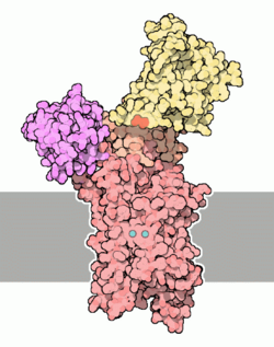
Ca2+ ATPase is a form of P-ATPase that transfers calcium after a muscle has contracted. The two kinds of calcium ATPase are:[2]
- Plasma membrane Ca2+ ATPase (PMCA)
- Sarcoplasmic reticulum Ca2+ ATPase (SERCA)
Plasma membrane Ca2+ ATPase (PMCA)

Plasma membrane Ca2+ ATPase (PMCA) is a transport protein in the plasma membrane of cells that serves to remove calcium (Ca2+) from the cell. It is vital for regulating the amount of Ca2+ within cells.[3] In fact, the PMCA is involved in removing Ca2+ from all eukaryotic cells.[4] There is a very large transmembrane electrochemical gradient of Ca2+ driving the entry of the ion into cells, yet it is very important for cells to maintain low concentrations of Ca2+ for proper cell signalling; thus it is necessary for the cell to employ ion pumps to remove the Ca2+.[5] The PMCA and the sodium calcium exchanger (NCX) are together the main regulators of intracellular Ca2+ concentrations.[4] Since it transports Ca2+ into the extracellular space, the PMCA is also an important regulator of the calcium concentration in the extracellular space.[6]
The PMCA belongs to a family of P-type primary ion transport ATPases that form an aspartyl phosphate intermediate.[4]
The PMCA is expressed in a variety of tissues, including the brain.[7]
Sarcoendoplasmic Reticulum Ca2+ ATPase (SERCA)
In myocytes (muscle cells) Ca2+ is normally sequestered (isolated) in a specialized form of endoplasmic reticulum (ER) called sarcoplasmic reticulum (SR). It is a Ca2+ ATPase that transfers Ca2+ from the cytosol of the cell to the lumen of the SR at the expense of ATP hydrolysis during muscle relaxation. In the skeletal muscles the calcium pump in the sarcoplasmic reticulum membrane works in harmony with similar calcium pumps in the plasma membrane. This ensures that the cytosolic concentration of free calcium in resting muscle is below 0.1µM. The sarcoplasmic and endoplasmic reticulum calcium pumps are closely related in structure and mechanism, and both are inhibited by the tumor-promoting agent thapsigargin, which does not affect the plasma membrane Ca2+ pumps.
See also
References
- ↑ PDB Molecule of the Month Calcium pump
- ↑ nlm.nih.gov
- ↑ "Expression of plasma membrane Ca2+ ATPase family members and associated synaptic proteins in acute and cultured organotypic hippocampal slices from rat". Brain Research. Developmental Brain Research 152 (2): 129–36. September 2004. doi:10.1016/j.devbrainres.2004.06.004. PMID 15351500.
- ↑ 4.0 4.1 4.2 "Role of alternative splicing in generating isoform diversity among plasma membrane calcium pumps". Physiological Reviews (American Physiological Society) 81 (1): 21–50. January 2001. doi:10.1152/physrev.2001.81.1.21. PMID 11152753. http://physrev.physiology.org/cgi/content/full/81/1/21?ijkey=febc5196952b9604d7293e211729df5e16861525.
- ↑ "Calcium pump of the plasma membrane". Physiological Reviews 71 (1): 129–53. January 1991. doi:10.1152/physrev.1991.71.1.129. PMID 1986387. http://physrev.physiology.org/cgi/reprint/71/1/129?ijkey=07dce5327b657cc65fb6152751c728391912b332&keytype2=tf_ipsecsha.
- ↑ "Expression and immunolocalization of plasma membrane calcium ATPase isoforms in human corneal epithelium". Molecular Vision 11: 169–78. March 2005. PMID 15765049. http://www.molvis.org/molvis/v11/a19/.
- ↑ "Presynaptic plasma membrane Ca2+ ATPase isoform 2a regulates excitatory synaptic transmission in rat hippocampal CA3". The Journal of Physiology 579 (Pt 1): 85–99. February 2007. doi:10.1113/jphysiol.2006.123901. 17170045. PMID 17170045. PMC 2075377. http://jp.physoc.org/cgi/rapidpdf/jphysiol.2006.123901v1.
External links
- Calcium+ATPase at the US National Library of Medicine Medical Subject Headings (MeSH)
- EC 7.2.2.10
- Overview at utoronto.ca
- Cation transporting ATPase family in Pfam
 |
