Biology:Phosphoglycerate dehydrogenase
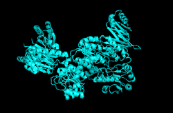
 Generic protein structure example |
| phosphoglycerate dehydrogenase | |||||||||
|---|---|---|---|---|---|---|---|---|---|
| Identifiers | |||||||||
| EC number | 1.1.1.95 | ||||||||
| Databases | |||||||||
| IntEnz | IntEnz view | ||||||||
| BRENDA | BRENDA entry | ||||||||
| ExPASy | NiceZyme view | ||||||||
| KEGG | KEGG entry | ||||||||
| MetaCyc | metabolic pathway | ||||||||
| PRIAM | profile | ||||||||
| PDB structures | RCSB PDB PDBe PDBsum | ||||||||
| |||||||||
In enzymology, D-3-phosphoglycerate dehydrogenase (PHGDH) (EC 1.1.1.95) is an enzyme that primarily catalyzes the chemical reactions
- 3-phospho-D-glycerate + NAD+ [math]\displaystyle{ \rightleftharpoons }[/math] 3-phosphonooxypyruvate + NADH + H+
- 2-hydroxyglutarate + NAD+ [math]\displaystyle{ \rightleftharpoons }[/math] 2-oxoglutarate + NADH + H+
Thus, in the first case, the two substrates of this enzyme are 3-phospho-D-glycerate and NAD+, whereas its 3 products are 3-phosphohydroxypyruvate, NADH, and H+; in the second case, the two substrates of this enzyme are 2-hydroxyglutarate and NAD+, whereas its 3 products are 2-oxoglutarate, NADH, and H+.
This enzyme belongs to the family of oxidoreductases, specifically those acting on the CH-OH group of donor with NAD+ or NADP+ as acceptor.
The most widely studied variants of PHGDH are from the E. coli and M. tuberculosis genomes.[1] In humans, this enzyme is encoded by the PHGDH gene.[2]
Function
3-Phosphoglycerate dehydrogenase catalyzes the transition of 3-phosphoglycerate into 3-phosphohydroxypyruvate, which is the committed step in the phosphorylated pathway of L-serine biosynthesis. It is also essential in cysteine and glycine synthesis, which lie further downstream.[3] This pathway represents the only way to synthesize serine in most organisms except plants, which uniquely possess multiple synthetic pathways. Nonetheless, the phosphorylated pathway that PHGDH participates in is still suspected to have an essential role in serine synthesis used in the developmental signaling of plants.[4][5]
Because of serine and glycine's role as neurotrophic factors in the developing brain, PHGDH has been shown to have high expression in glial and astrocyte cells during neural development.[6]
Mechanism and regulation
3-phosphoglycerate dehydrogenase works via an induced fit mechanism to catalyze the transfer of a hydride from the substrate to NAD+, a required cofactor. In its active conformation, the enzyme's active site has multiple cationic residues that likely stabilize the transition state of the reaction between the negatively charged substrate and NAD+. The positioning is such that the substrate's alpha carbon and the C4 of the nicotinamide ring are brought into a proximity that facilitates the hydride transfer producing NADH and the oxidized substrate.[1][7]
PHGDH is allosterically regulated by its downstream product, L-serine. This feedback inhibition is understandable considering that 3-phosphoglycerate is an intermediate in the glycolytic pathway. Given that PHGDH represents the committed step in the production of serine in the cell, flux through the pathway must be carefully controlled.
L-serine binding has been shown to exhibit cooperative behavior. Mutants that decreased this cooperativity also increased in sensitivity to serine's allosteric inhibition, suggesting a separation of the chemical mechanisms that result in allosteric binding cooperativity and active site inhibition.[8] The mechanism of inhibition is Vmax type, indicating that serine affects the reaction rate rather than the binding affinity of the active site.[7][9]
Although L-serine's allosteric effects are usually the focus of regulatory investigation, it has been noted that in some variants of the enzyme, 3-phosphoglycerate dehydrogenase is inhibited at separate positively charged allosteric site by high concentrations of its own substrate.[1][10]
Structure
3-Phosphoglycerate dehydrogenase is a tetramer, composed of four identical, asymmetric subunits. At any time, only a maximum of two adjacent subunits present a catalytically active site; the other two are forced into an inactive conformation. This results in half-of-the-sites activity with regard to both active and allosteric sites, meaning that only the two sites of the active subunits must be bound for essentially maximal effect with regard to catalysis and inhibition respectively.[11] There is some evidence that further inhibition occurs with the binding of the third and fourth serine molecules, but it is relatively minimal.[9]
The subunits from the E. coli PHGDH have three distinct domains, whereas those from M. tuberculosis have four. It is noted that the human enzyme more closely resembles that of M. tuberculosis, including the site for allosteric substrate inhibition. Concretely, three general types of PHGDH have been proposed: Type I, II, and III. Type III has two distinct domains, lacks both allosteric sites, and is found in various unicellular organisms. Type II has serine binding sites and encompasses the well-studied E. coli PHGDH. Type I possesses both the serine and substrate allosteric binding sites and encompasses M. tuberculosis and mammalian PHGDHs.[1]
The regulation of catalytic activity is thought to be a result of the movement of rigid domains about flexible “hinges.” When the substrate binds to the open active site, the hinge rotates and closes the cleft. Allosteric inhibition thus likely works by locking the hinge in a state that produces the open active site cleft.[9][12]
The variant from M. tuberculosis also exhibits an uncommon dual pH optimum for catalytic activity.[10]
Evolution
3-Phosphoglycerate dehydrogenase possesses less than 20% homology to other NAD-dependent oxidoreductases and exhibits significant variance between species. There does appear to be conservation in specific binding domain residues, but there is still some variation in the positively charged active site residues between variants. For example, Type III PHGDH enzymes can be broken down into two subclasses where the key histidine residue is replaced with a lysine residue.[1][13]
Disease relevance
Homozygous or compound heterozygous mutations in 3-phosphoglycerate dehydrogenase cause Neu-Laxova syndrome[14][15] and phosphoglycerate dehydrogenase deficiency.[16] In addition significantly shortening lifespan, PHGDH deficiencies are known to cause congenital microcephaly, psychomotor retardation, and intractable seizures in both humans and rats, presumably due to the essential signaling within the nervous system that serine, glycine, and other downstream molecules are intimately involved with. Treatment typically involves oral supplementation of serine and glycine and has been shown most effective when started in utero via oral ingestion by the mother.[17][18]
Mutations that result in increased PHGDH activity are also associated with increased risk of oncogenesis, including certain breast cancers.[19] This finding suggests that pathways providing an outlet for diverting carbon out of glycolysis may be beneficial for rapid cell growth.[20]
It has been reported that PHGDH can also catalyze the conversion of alpha-ketoglutarate to 2-hydroxyglutaric acid in certain variants. Thus, a mutation in the enzyme is hypothesized to contribute to 2-hydroxyglutaric aciduria in humans, although there is debate as to whether or not this catalysis is shared by human PHGDH.[1][21]
References
- ↑ 1.0 1.1 1.2 1.3 1.4 1.5 "Contrasting catalytic and allosteric mechanisms for phosphoglycerate dehydrogenases". Archives of Biochemistry and Biophysics 519 (2): 175–85. Mar 2012. doi:10.1016/j.abb.2011.10.005. PMID 22023909.
- ↑ "PHGDH phosphoglycerate dehydrogenase [Homo sapiens (human) - Gene - NCBI"]. https://www.ncbi.nlm.nih.gov/gene?Db=gene&Cmd=ShowDetailView&TermToSearch=26227.
- ↑ "MetaCyc L-serine biosynthesis". http://biocyc.org/META/NEW-IMAGE?type=PATHWAY&object=SERSYN-PWY.
- ↑ "Serine in plants: biosynthesis, metabolism, and functions". Trends in Plant Science 19 (9): 564–9. Sep 2014. doi:10.1016/j.tplants.2014.06.003. PMID 24999240.
- ↑ "Regulation of serine biosynthesis in Arabidopsis. Crucial role of plastidic 3-phosphoglycerate dehydrogenase in non-photosynthetic tissues". The Journal of Biological Chemistry 274 (1): 397–402. Jan 1999. doi:10.1074/jbc.274.1.397. PMID 9867856.
- ↑ "3-Phosphoglycerate dehydrogenase, a key enzyme for l-serine biosynthesis, is preferentially expressed in the radial glia/astrocyte lineage and olfactory ensheathing glia in the mouse brain". The Journal of Neuroscience 21 (19): 7691–704. Oct 2001. PMID 11567059.
- ↑ 7.0 7.1 "The contribution of adjacent subunits to the active sites of D-3-phosphoglycerate dehydrogenase". The Journal of Biological Chemistry 274 (9): 5357–61. Feb 1999. doi:10.1074/jbc.274.9.5357. PMID 10026144.
- ↑ "Specific interactions at the regulatory domain-substrate binding domain interface influence the cooperativity of inhibition and effector binding in Escherichia coli D-3-phosphoglycerate dehydrogenase". The Journal of Biological Chemistry 276 (2): 1078–83. Jan 2001. doi:10.1074/jbc.M007512200. PMID 11050089.
- ↑ 9.0 9.1 9.2 "A model for the regulation of D-3-phosphoglycerate dehydrogenase, a Vmax-type allosteric enzyme". Protein Science 5 (1): 34–41. Jan 1996. doi:10.1002/pro.5560050105. PMID 8771194.
- ↑ 10.0 10.1 "A novel mechanism for substrate inhibition in Mycobacterium tuberculosis D-3-phosphoglycerate dehydrogenase". The Journal of Biological Chemistry 282 (43): 31517–24. Oct 2007. doi:10.1074/jbc.M704032200. PMID 17761677.
- ↑ "Quantitative relationships of site to site interaction in Escherichia coli D-3-phosphoglycerate dehydrogenase revealed by asymmetric hybrid tetramers". The Journal of Biological Chemistry 279 (14): 13452–60. Apr 2004. doi:10.1074/jbc.M313593200. PMID 14718528.
- ↑ "The mechanism of velocity modulated allosteric regulation in D-3-phosphoglycerate dehydrogenase. Cross-linking adjacent regulatory domains with engineered disulfides mimics effector binding". The Journal of Biological Chemistry 271 (22): 13013–7. May 1996. PMID 8662776.
- ↑ "The nucleotide sequence of the serA gene of Escherichia coli and the amino acid sequence of the encoded protein, D-3-phosphoglycerate dehydrogenase". The Journal of Biological Chemistry 261 (26): 12179–83. Sep 1986. PMID 3017965.
- ↑ "Neu-Laxova syndrome, an inborn error of serine metabolism, is caused by mutations in PHGDH". American Journal of Human Genetics 94 (6): 898–904. Jun 2014. doi:10.1016/j.ajhg.2014.04.015. PMID 24836451.
- ↑ "Neu-Laxova syndrome is a heterogeneous metabolic disorder caused by defects in enzymes of the L-serine biosynthesis pathway". American Journal of Human Genetics 95 (3): 285–93. Sep 2014. doi:10.1016/j.ajhg.2014.07.012. PMID 25152457.
- ↑ "3-Phosphoglycerate dehydrogenase deficiency: an inborn error of serine biosynthesis". Archives of Disease in Childhood 74 (6): 542–5. Jun 1996. doi:10.1136/adc.74.6.542. PMID 8758134.
- ↑ "Beneficial effects of L-serine and glycine in the management of seizures in 3-phosphoglycerate dehydrogenase deficiency". Annals of Neurology 44 (2): 261–5. Aug 1998. doi:10.1002/ana.410440219. PMID 9708551.
- ↑ "Prenatal and early postnatal treatment in 3-phosphoglycerate-dehydrogenase deficiency". Lancet 364 (9452): 2221–2. 2004-12-18. doi:10.1016/S0140-6736(04)17596-X. PMID 15610810.
- ↑ "Functional genomics reveal that the serine synthesis pathway is essential in breast cancer". Nature 476 (7360): 346–50. Aug 2011. doi:10.1038/nature10350. PMID 21760589.
- ↑ "Phosphoglycerate dehydrogenase diverts glycolytic flux and contributes to oncogenesis". Nature Genetics 43 (9): 869–74. Sep 2011. doi:10.1038/ng.890. PMID 21804546. PMC 3677549. http://dspace.mit.edu/bitstream/1721.1/76302/1/PHGDH_NatGen_FINAL2.0.pdf.
- ↑ "A novel alpha-ketoglutarate reductase activity of the serA-encoded 3-phosphoglycerate dehydrogenase of Escherichia coli K-12 and its possible implications for human 2-hydroxyglutaric aciduria". Journal of Bacteriology 178 (1): 232–9. Jan 1996. PMID 8550422.
Further reading
- "A systematic analysis of human CHMP protein interactions: additional MIT domain-containing proteins bind to multiple components of the human ESCRT III complex". Genomics 88 (3): 333–46. Sep 2006. doi:10.1016/j.ygeno.2006.04.003. PMID 16730941.
- "Proteomic analysis of SUMO4 substrates in HEK293 cells under serum starvation-induced stress". Biochemical and Biophysical Research Communications 337 (4): 1308–18. Dec 2005. doi:10.1016/j.bbrc.2005.09.191. PMID 16236267.
- "V490M, a common mutation in 3-phosphoglycerate dehydrogenase deficiency, causes enzyme deficiency by decreasing the yield of mature enzyme". The Journal of Biological Chemistry 277 (9): 7136–43. Mar 2002. doi:10.1074/jbc.M111419200. PMID 11751922.
- "Molecular characterization of 3-phosphoglycerate dehydrogenase deficiency--a neurometabolic disorder associated with reduced L-serine biosynthesis". American Journal of Human Genetics 67 (6): 1389–99. Dec 2000. doi:10.1086/316886. PMID 11055895.
- "3-phosphoglycerate dehydrogenase deficiency in a patient with West syndrome". Developmental Medicine and Child Neurology 42 (9): 629–33. Sep 2000. doi:10.1017/S0012162200001171. PMID 11034457.
- "Assignment of human 3-phosphoglycerate dehydrogenase (PHGDH) to human chromosome band 1p12 by fluorescence in situ hybridization". Cytogenetics and Cell Genetics 89 (1-2): 6–7. 2000. doi:10.1159/000015577. PMID 10894924.
- "Nucleotide sequence and differential expression of the human 3-phosphoglycerate dehydrogenase gene". Gene 245 (1): 193–201. Mar 2000. doi:10.1016/S0378-1119(00)00009-3. PMID 10713460.
- "3-Phosphoglycerate dehydrogenase deficiency: an inborn error of serine biosynthesis". Archives of Disease in Childhood 74 (6): 542–5. Jun 1996. doi:10.1136/adc.74.6.542. PMID 8758134.
External links
- Overview of all the structural information available in the PDB for UniProt: O43175 (D-3-phosphoglycerate dehydrogenase) at the PDBe-KB.


