Biology:Rhizaria
| Rhizaria Temporal range: 650 Mya[1] (Neoproterozoic) - Present
| |
|---|---|

| |
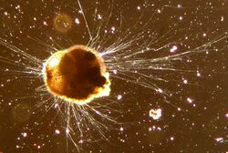
| Ammonia tepida (Foraminifera) | |
| Scientific classification | |
| Domain: | Eukaryota |
| Clade: | Diaphoretickes |
| Clade: | TSAR |
| Clade: | SAR |
| Clade: | Rhizaria Cavalier-Smith, 2002 |
| Phyla | |
The Rhizaria are a diverse and species-rich supergroup of mostly unicellular[2] eukaryotes.[3] Except for the Chlorarachniophytes and three species in the genus Paulinella in the phylum Cercozoa, they are all non-photosynthethic, but many foraminifera and radiolaria have a symbiotic relationship with unicellular algae.[4] A multicellular form, Guttulinopsis vulgaris, a cellular slime mold, has been described.[5] This group was used by Cavalier-Smith in 2002, although the term "Rhizaria" had been long used for clades within the currently recognized taxon. Being described mainly from rDNA sequences, they vary considerably in form, having no clear morphological distinctive characters (synapomorphies), but for the most part they are amoeboids with filose, reticulose, or microtubule-supported pseudopods. In the absence of an apomorphy, the group is ill-defined, and its composition has been very fluid. Some Rhizaria possess mineral exoskeletons (thecae or loricas), which are in different clades within Rhizaria made out of opal (SiO
2), celestite (SrSO
4), or calcite (CaCO
3). Certain species can attain sizes of more than a centimeter with some species being able to form cylindrical colonies approximately 1 cm in diameter and greater than 1 m in length. They feed by capturing and engulfing prey with the extensions of their pseudopodia; forms that are symbiotic with unicellular algae contribute significantly to the total primary production of the ocean.[6]
Groups
The three main groups of Rhizaria are:[7]
- Cercozoa – various amoebae and flagellates, usually with filose pseudopods and common in soil
- Foraminifera – amoeboids with reticulose pseudopods, common as marine benthos
- Radiolaria – amoeboids with axopods, common as marine plankton
A few other groups may be included in the Cercozoa, but some trees appear closer to the Foraminifera. These are the Phytomyxea and Ascetosporea, parasites of plants and animals, respectively, and the peculiar amoeba Gromia. The different groups of Rhizaria are considered close relatives based mainly on genetic similarities, and have been regarded as an extension of the Cercozoa. The name Rhizaria for the expanded group was introduced by Cavalier-Smith in 2002,[8] who also included the centrohelids and Apusozoa.
A noteworthy order that belongs to Ascetosporea is the Mikrocytida.[9] These are parasites of oysters. This includes the causative agent of Denman Island Disease, Mikrocytos mackini a small (2−3 μm diameter) amitochondriate protistan.[10]
Evolutionary relationships
Rhizaria are part of the SAR supergroup (Stramenopiles, Alveolates, Rhizaria), a grouping that had been presaged in 1993 through a study of mitochondrial morphologies.[11] SAR is currently placed in the Diaphoretickes along with Archaeplastida, Cryptista, Haptista, and several minor clades.
Historically, many rhizarians were considered animals because of their motility and heterotrophy. However, when a simple animal-plant dichotomy was superseded by a recognition of additional kingdoms, taxonomists generally placed amoebae in the kingdom Protista. When scientists began examining the evolutionary relationships among eukaryotes in the 1970s, it became clear that the kingdom Protista was paraphyletic. Rhizaria appear to share a common ancestor with Stramenopiles and Alveolates forming part of the SAR super assemblage.[12] Rhizaria has been supported by molecular phylogenetic studies as a monophyletic group.[13] Biosynthesis of 24-isopropyl cholestane precursors in various rhizaria[14] suggests a relevant ecological role already during the Ediacaran.
Phylogeny
Rhizaria is a monophyletic group composed of two sister phyla: Cercozoa and Retaria. Subsequently, Cercozoa and Retaria are also monophyletic.[15][16] The following cladogram depicts the evolutionary relationships between all rhizarian classes, and is made after the works of Cavalier-Smith et al. (2018),[1] and Irwin et al. (2019).[17]
| SAR Supergroup |
| ||||||||||||||||||||||||||||||||||||||||||||||||||||||||||||||||||||||||||||||||||||||||||
Sexual cycle
Complete sexual life cycles have been demonstrated for two lineages (Foraminifera and Gromia) and direct evidence for karyogamy or meiosis has been observed in five lineages (Euglyphida, Thecofilosea, Chlorarachniophyta, Plasmodiophorida and Phaeodarea).[18] In particular, the Foramanifera are marine amoebae that are defined by a dynamic network of pseudopodia, and the production of intricate shells.[18] These amoeba have complex sexual life cycles with meiosis and gamete production occurring at separate stages.[18]
References
- ↑ 1.0 1.1 Cavalier-Smith, Thomas; Chao, Ema E .; Lewis, Rhodri (April 2018). "Multigene phylogeny and cell evolution of chromist infrakingdom Rhizaria: contrasting cell organisation of sister phyla Cercozoa and Retaria". Protoplasma 255 (5): 1517–1574. doi:10.1007/s00709-018-1241-1. PMID 29666938.
- ↑ Taylor, Christopher (2004). "Rhizaria". http://www.palaeos.com/Eukarya/Units/Rhizaria/Rhizaria.html.
- ↑ Nikolaev, Sergey I.; Berney, Cédric; Fahrni, José F. et al. (May 2004). "The twilight of Heliozoa and rise of Rhizaria, an emerging supergroup of amoeboid eukaryotes". PNAS 101 (21): 8066–71. doi:10.1073/pnas.0308602101. PMID 15148395.
- ↑ Gast, Rebecca J.; Caron, David A. (2001-10-01). "Photosymbiotic associations in planktonic foraminifera and radiolaria". Hydrobiologia 461 (1): 1–7. doi:10.1023/A:1012710909023.
- ↑ Brown, Matthew W.; Kolisko, Martin; Silberman, Jeffrey D.; Roger, Andrew J. (2012). "Aggregative Multicellularity Evolved Independently in the Eukaryotic Supergroup Rhizaria". Current Biology 22 (12): 1123–1127. doi:10.1016/j.cub.2012.04.021.
- ↑ Caron, D. (2016). The rise of Rhizaria. Nature (London), 532(7600), 444–445. https://doi.org/10.1038/nature17892
- ↑ "Global eukaryote phylogeny: Combined small- and large-subunit ribosomal DNA trees support monophyly of Rhizaria, Retaria and Excavata". Mol. Phylogenet. Evol. 44 (1): 255–66. July 2007. doi:10.1016/j.ympev.2006.11.001. PMID 17174576.
- ↑ Cavalier-Smith, Thomas (2002). "The phagotrophic origin of eukaryotes and phylogenetic classification of Protozoa". International Journal of Systematic and Evolutionary Microbiology 52 (2): 297–354. doi:10.1099/00207713-52-2-297. ISSN 1466-5026. PMID 11931142. http://ijs.sgmjournals.org/cgi/content/abstract/52/2/297. Retrieved 2007-06-08.
- ↑ Hartikainen, H.; Stentiford, G.D.; Bateman, K.S.; Berney, C.; Feist, S.W.; Longshaw, M.; Okamura, B.; Stone, D. et al. (2014). "Mikrocytids are a broadly distributed and divergent radiation of parasites in aquatic invertebrates". Curr Biol 24 (7): 807–12. doi:10.1016/j.cub.2014.02.033. PMID 24656829. https://nhm.openrepository.com/bitstream/10141/622246/1/Mikrocytids_CurrBiol_2014.pdf.
- ↑ Hine, P.M.; Bower, S.M.; Meyer, G.R.; Cochennec-Laureau, N.; Berthe, F.C.J. (2001). "Ultrastructure of Mikrocytos mackini, the cause of Denman Island disease in oysters Crassostrea spp. and Ostrea spp. in British Columbia, Canada". Diseases of Aquatic Organisms 45 (3): 215–227. doi:10.3354/dao045215. ISSN 0177-5103. PMID 11558731. http://www.int-res.com/abstracts/dao/v45/n3/p215-227/.
- ↑ Seravin LN. Osnovnye tipy i formy tonkogo stroeniia krist mitokhondriĭ: stepen' ikh évoliutsionnoĭ stabil'nosti (sposobnost' k morfologicheskim transformatsiiam) [The basic types and forms of the fine structure of mitochondrial cristae: the degree of their evolutionary stability (capacity for morphological transformations)]. Tsitologiia. 1993;35(4):3-34. Russian. PMID 8328023.
- ↑ Burki, F.; Shalchian-Tabrizi, K.; Minge, M.; Skjaeveland, A.; Nikolaev, S.I.; Jakobsen, K.S.; Pawlowski, J. (2007). Butler, Geraldine. ed. "Phylogenomics Reshuffles the Eukaryotic Supergroups". PLoS ONE 2 (8): e790–. doi:10.1371/journal.pone.0000790. PMID 17726520. Bibcode: 2007PLoSO...2..790B.
- ↑ Burki, Fabien; Shalchian-Tabrizi, Kamran; Pawlowski, Jan (August 23, 2008). "Phylogenomics reveals a new 'megagroup' including most photosynthetic eukaryotes". Biology Letters 4 (4): 366–369. doi:10.1098/rsbl.2008.0224. PMID 18522922.
- ↑ Hallmann, Christian; Stuhr, Marleen; Kucera, Michal et al. (2019-03-04). "Putative sponge biomarkers in unicellular Rhizaria question an early rise of animals". Nature Ecology & Evolution 3 (4): 577–581. doi:10.1038/s41559-019-0806-5. PMID 30833757.
- ↑ Bass, D. et al. (February 2009). "Phylogeny of Novel Naked Filose and Reticulose Cercozoa: Granofilosea cl. n. and Proteomyxidea Revised". Protist 160 (1): 75–109. doi:10.1016/j.protis.2008.07.002. PMID 18952499.
- ↑ >Howe, Alexis T.; Bass, David; Scoble, Josephine M. et al. (2011). "Novel Cultured Protists Identify Deep-branching Environmental DNA Clades of Cercozoa: New Genera Tremula, Micrometopion, Minimassisteria, Nudifila, Peregrinia". Protist 162 (2): 332–372. doi:10.1016/j.protis.2010.10.002. PMID 21295519.
- ↑ Irwin, Nicholas A. T.; Tikhonenkov, Denis V.; Hehenberger, Elisabeth et al. (2019-01-01). "Phylogenomics supports the monophyly of the Cercozoa". Molecular Phylogenetics and Evolution 130: 416–423. doi:10.1016/j.ympev.2018.09.004. ISSN 1055-7903. PMID 30318266.
- ↑ 18.0 18.1 18.2 Lahr DJ, Parfrey LW, Mitchell EA, Katz LA, Lara E. The chastity of amoebae: re-evaluating evidence for sex in amoeboid organisms. Proc Biol Sci. 2011 Jul 22;278(1715):2081-90. doi: 10.1098/rspb.2011.0289. Epub 2011 Mar 23. PMID: 21429931; PMCID: PMC3107637
External links
- Rhizaria at UniEuk Taxonomy Map
- Molecular Phylogeny of Amoeboid Protists - Tree of Rhizaria
- Tree of Life Eukaryota
Wikidata ☰ Q855740 entry
 |


