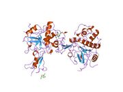
Biology:Cathepsin L1
 Generic protein structure example |
Cathepsin L1 is a protein that in humans is encoded by the CTSL1 gene.[1][2][3] The protein is a cysteine cathepsin, a lysosomal cysteine protease that plays a major role in intracellular protein catabolism.[4][5][6][7]
Function
Cathepsin L1 is a member of the Peptidase C1 (cathepsin) MEROPS family, which plays an important role in diverse processes including normal lysosome mediated protein turnover, antigen and proprotein processing, and apoptosis.[8] Its substrates include collagen and elastin, as well as alpha-1 protease inhibitor, a major controlling element of neutrophil elastase activity. The encoded protein has been implicated in several pathologic processes, including myofibril necrosis in myopathies and in myocardial ischemia, and in the renal tubular response to proteinuria. This protein, which is a member of the peptidase C1 family, is a dimer composed of disulfide-linked heavy and light chains, both produced from a single protein precursor. At least two transcript variants encoding the same protein have been found for this gene.[3]
Viral entry
Cleavage of the SARS-CoV-2 S2 spike protein required for viral entry into cells can be accomplished by proteases TMPRSS2 located on the cell membrane, or by cathepsins (primarily cathepsin L) in endolysosomes.[9] Hydroxychloroquine inhibits the action of cathepsin L in endolysosomes, but because cathepsin L cleavage is minor compared to TMPRSS2 cleavage, hydroxychloroquine does little to inhibit SARS-CoV-2 infection.[9]
Inflammation
Although Cathepsin L is usually characterized as a lysosomal protease, it can be secreted, resulting in pathological inflammation.[10] Cathepsin L and other cysteine cathepsins tend to be secreted by macrophages and other tissue-invading immune cells when causing pathological inflammation.[11]
Interactions
CTSL1 has been shown to interact with Cystatin A.[12][13]
Distribution
Cathepsin L has been reported in many organisms including fish,[14] birds, mammals, and sponges.[15]
See also
- Cathepsin L2 (also known as cathepsin V)
References
- ↑ "Cloning, genomic organization, and chromosomal localization of human cathepsin L". J Biol Chem 268 (2): 1039–45. Feb 1993. doi:10.1016/S0021-9258(18)54038-2. PMID 8419312.
- ↑ "Complete nucleotide and deduced amino acid sequences of human and murine preprocathepsin L. An abundant transcript induced by transformation of fibroblasts". J Clin Invest 81 (5): 1621–9. Jun 1988. doi:10.1172/JCI113497. PMID 2835398.
- ↑ 3.0 3.1 "Entrez Gene: CTSL1 cathepsin L1". https://www.ncbi.nlm.nih.gov/sites/entrez?Db=gene&Cmd=ShowDetailView&TermToSearch=1514.
- ↑ "Cathepsin B, cathepsin H, and cathepsin L". Proteolytic Enzymes, Part C. Methods in Enzymology. 80 Pt C. 1981. pp. 535–561. doi:10.1016/s0076-6879(81)80043-2. ISBN 9780121819804.
- ↑ "Lysosomal cysteine proteinases". ISI Atlas of Science. Biochemistry 1: 256–260. 1988.
- ↑ "Complete nucleotide and deduced amino acid sequences of human and murine preprocathepsin L. An abundant transcript induced by transformation of fibroblasts". The Journal of Clinical Investigation 81 (5): 1621–1629. May 1988. doi:10.1172/JCI113497. PMID 2835398.
- ↑ "Active center differences between cathepsins L and B: the S1 binding region". FEBS Letters 228 (1): 128–130. February 1988. doi:10.1016/0014-5793(88)80600-8. PMID 3342870.
- ↑ "Cysteine Peptidases of Mammals: Their Biological Roles and Potential Effects in the Oral Cavity and Other Tissues in Health and Disease". Critical Reviews in Oral Biology and Medicine 13 (3): 238–75. 2002. doi:10.1177/154411130201300304. PMID 12090464.
- ↑ 9.0 9.1 "Mechanisms of SARS-CoV-2 entry into cells". Nature Reviews. Molecular Cell Biology 23 (1): 3–20. January 2022. doi:10.1038/s41580-021-00418-x. PMID 34611326.
- ↑ "Cathepsin L in COVID-19: From Pharmacological Evidences to Genetics". Frontiers in Cellular and Infection Microbiology 10: 589505. 2022. doi:10.3389/fcimb.2020.589505. PMID 33364201.
- ↑ "Cathepsin L, transmembrane peptidase/serine subfamily member 2/4, and other host proteases in COVID-19 pathogenesis - with impact on gastrointestinal tract". World Journal of Gastroenterology 27 (39): 6590–6600. October 2021. doi:10.3748/wjg.v27.i39.6590. PMID 34754154.
- ↑ Majerle, Andreja; Jerala Roman (Sep 2003). "Protein inhibitors form complexes with procathepsin L and augment cleavage of the propeptide". Arch. Biochem. Biophys. 417 (1): 53–8. doi:10.1016/S0003-9861(03)00319-9. ISSN 0003-9861. PMID 12921779.
- ↑ Estrada, S; Nycander M; Hill N J; Craven C J; Waltho J P; Björk I (May 1998). "The role of Gly-4 of human cystatin A (stefin A) in the binding of target proteinases. Characterization by kinetic and equilibrium methods of the interactions of cystatin A Gly-4 mutants with papain, cathepsin B, and cathepsin L". Biochemistry 37 (20): 7551–60. doi:10.1021/bi980026r. ISSN 0006-2960. PMID 9585570.
- ↑ "A murrel cysteine protease, cathepsin L: bioinformatics characterization, gene expression and proteolytic activity". Biologia 39 (3): 395–406. 2014. doi:10.2478/s11756-013-0326-8.
- ↑ "Human cathepsin L rescues the neurodegeneration and lethality in cathepsin B/L double-deficient mice". Biological Chemistry 387 (7): 885–91. July 2006. doi:10.1515/BC.2006.112. PMID 16913838. https://digital.library.unt.edu/ark:/67531/metadc896009/.
Further reading
- "Effect of delta 9-tetrahydrocannabinol (THC) on female reproductive function.". Advances in the Biosciences 22-23: 449–67. 1980. doi:10.1016/b978-0-08-023759-6.50040-8. ISBN 9780080237596. PMID 116880.
- "Effective activation of the proenzyme form of the urokinase-type plasminogen activator (pro-uPA) by the cysteine protease cathepsin L.". FEBS Lett. 297 (1–2): 112–8. 1992. doi:10.1016/0014-5793(92)80339-I. PMID 1551416.
- "Thyroglobulin processing by thyroidal proteases. Major sites of cleavage by cathepsins B, D, and L.". J. Biol. Chem. 266 (30): 20198–204. 1991. doi:10.1016/S0021-9258(18)54909-7. PMID 1939080.
- "Comparison of cathepsin L synthesized by normal and transformed cells at the gene, message, protein, and oligosaccharide levels.". Arch. Biochem. Biophys. 283 (2): 447–57. 1991. doi:10.1016/0003-9861(90)90666-M. PMID 2275556.
- "Amino acid sequences of the human kidney cathepsins H and L.". FEBS Lett. 228 (2): 341–5. 1988. doi:10.1016/0014-5793(88)80028-0. PMID 3342889.
- "Isolation and sequence of a cDNA for human pro-(cathepsin L).". Biochem. J. 253 (1): 303–6. 1988. doi:10.1042/bj2530303. PMID 3421948.
- "Cathepsin L inactivates alpha 1-proteinase inhibitor by cleavage in the reactive site region.". J. Biol. Chem. 261 (31): 14748–51. 1986. doi:10.1016/S0021-9258(18)66935-2. PMID 3490478.
- "The N-terminal amino acid sequences of the heavy and light chains of human cathepsin L. Relationship to a cDNA clone for a major cysteine proteinase from a mouse macrophage cell line.". Biochem. J. 240 (2): 373–7. 1987. doi:10.1042/bj2400373. PMID 3545185.
- "The major ras induced protein in NIH3T3 cells is cathepsin L.". Nucleic Acids Res. 15 (7): 3186. 1987. doi:10.1093/nar/15.7.3186. PMID 3550705.
- "Action of rat liver cathepsin L on glucagon.". Acta Biol. Med. Ger. 40 (9): 1139–43. 1982. PMID 7340337.
- "Major histocompatibility complex class II-associated p41 invariant chain fragment is a strong inhibitor of lysosomal cathepsin L.". J. Exp. Med. 183 (4): 1331–8. 1996. doi:10.1084/jem.183.4.1331. PMID 8666891.
- "Structure of human procathepsin L reveals the molecular basis of inhibition by the prosegment.". EMBO J. 15 (20): 5492–503. 1996. doi:10.1002/j.1460-2075.1996.tb00934.x. PMID 8896443.
- "Identification of peptide fragments generated by digestion of bovine and human osteocalcin with the lysosomal proteinases cathepsin B, D, L, H, and S.". J. Bone Miner. Res. 12 (3): 447–55. 1997. doi:10.1359/jbmr.1997.12.3.447. PMID 9076588.
- "The crystal structure of human cathepsin L complexed with E-64.". FEBS Lett. 407 (1): 47–50. 1997. doi:10.1016/S0014-5793(97)00216-0. PMID 9141479.
- "Autocatalytic processing of recombinant human procathepsin L. Contribution of both intermolecular and unimolecular events in the processing of procathepsin L in vitro.". J. Biol. Chem. 273 (8): 4478–84. 1998. doi:10.1074/jbc.273.8.4478. PMID 9468501.
- "Cross-class inhibition of the cysteine proteinases cathepsins K, L, and S by the serpin squamous cell carcinoma antigen 1: a kinetic analysis.". Biochemistry 37 (15): 5258–66. 1998. doi:10.1021/bi972521d. PMID 9548757.
- "The role of Gly-4 of human cystatin A (stefin A) in the binding of target proteinases. Characterization by kinetic and equilibrium methods of the interactions of cystatin A Gly-4 mutants with papain, cathepsin B, and cathepsin L.". Biochemistry 37 (20): 7551–60. 1998. doi:10.1021/bi980026r. PMID 9585570.
- "Leukocystatin, a new Class II cystatin expressed selectively by hematopoietic cells.". J. Biol. Chem. 273 (26): 16400–8. 1998. doi:10.1074/jbc.273.26.16400. PMID 9632704.
External links
- The MEROPS online database for peptidases and their inhibitors: C01.032
- Cathepsin+L at the US National Library of Medicine Medical Subject Headings (MeSH)
 |


