Biology:Phosphoenolpyruvate carboxykinase
| Phosphoenolpyruvate carboxykinase | |||||||||||
|---|---|---|---|---|---|---|---|---|---|---|---|
 PDB rendering based on 1khb. | |||||||||||
| Identifiers | |||||||||||
| Symbol | PEPCK | ||||||||||
| Pfam | PF00821 | ||||||||||
| InterPro | IPR008209 | ||||||||||
| PROSITE | PDOC00421 | ||||||||||
| SCOP2 | 1khf / SCOPe / SUPFAM | ||||||||||
| |||||||||||
| phosphoenolpyruvate carboxykinase 1 (soluble) | |
|---|---|
 Phosphoenolpyruvate carboxykinase (GTP, cytosolic) monomer, Human | |
| Identifiers | |
| Symbol | PCK1 |
| Alt. symbols | PEPCK-C |
| NCBI gene | 5105 |
| HGNC | 8724 |
| OMIM | 261680 |
| RefSeq | NM_002591 |
| Other data | |
| EC number | 4.1.1.32 |
| Locus | Chr. 20 q13.31 |
| phosphoenolpyruvate carboxykinase 2 (mitochondrial) | |
|---|---|
| Identifiers | |
| Symbol | PCK2 |
| Alt. symbols | PEPCK-M, PEPCK2 |
| NCBI gene | 5106 |
| HGNC | 8725 |
| OMIM | 261650 |
| RefSeq | NM_001018073 |
| Other data | |
| EC number | 4.1.1.32 |
| Locus | Chr. 14 q12 |
Phosphoenolpyruvate carboxykinase (EC 4.1.1.32, PEPCK) is an enzyme in the lyase family used in the metabolic pathway of gluconeogenesis. It converts oxaloacetate into phosphoenolpyruvate and carbon dioxide.[1][2][3]
It is found in two forms, cytosolic and mitochondrial.
Structure
In humans there are two isoforms of PEPCK; a cytosolic form (SwissProt P35558) and a mitochondrial isoform (SwissProt Q16822) which have 63.4% sequence identity. The cytosolic form is important in gluconeogenesis. However, there is a known transport mechanism to move PEP from the mitochondria to the cytosol, using specific membrane transport proteins.[4][5][6][7][8] PEP transport across the inner mitochondrial membrane involves the mitochondrial tricarboxylate transport protein and to a lesser extent the adenine nucleotide carrier. The possibility of a PEP/pyruvate transporter has also been put forward.[9]
X-ray structures of PEPCK provide insight into the structure and the mechanism of PEPCK enzymatic activity. The mitochondrial isoform of chicken liver PEPCK complexed with Mn2+, Mn2+-phosphoenolpyruvate (PEP), and Mn2+-GDP provides information about its structure and how this enzyme catalyzes reactions.[10] Delbaere et al. (2004) resolved PEPCK in E. coli and found the active site sitting between a C-terminal domain and an N-terminal domain. The active site was observed to be closed upon rotation of these domains.[11]
Phosphoryl groups are transferred during PEPCK action, which is likely facilitated by the eclipsed conformation of the phosphoryl groups when ATP is bound to PEPCK.[11]
Since the eclipsed formation is one that is high in energy, phosphoryl group transfer has a decreased energy of activation, meaning that the groups will transfer more readily. This transfer likely happens via a mechanism similar to SN2 displacement.[11]
In different species
PEPCK gene transcription occurs in many species, and the amino acid sequence of PEPCK is distinct for each species.
For example, its structure and its specificity differ in humans, Escherichia coli (E. coli), and the parasiteTrypanosoma cruzi.[12]
Mechanism
PEPCKase converts oxaloacetate into phosphoenolpyruvate and carbon dioxide.
-
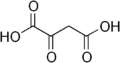 oxaloacetate
 -
 phosphoenolpyruvate


As PEPCK acts at the junction between glycolysis and the Krebs cycle, it causes decarboxylation of a C4 molecule, creating a C3 molecule. As the first committed step in gluconeogenesis, PEPCK decarboxylates and phosphorylates oxaloacetate (OAA) for its conversion to PEP, when GTP is present. As a phosphate is transferred, the reaction results in a GDP molecule.[10] When pyruvate kinase – the enzyme that normally catalyzes the reaction that converts PEP to pyruvate – is knocked out in mutants of Bacillus subtilis, PEPCK participates in one of the replacement anaplerotic reactions, working in the reverse direction of its normal function, converting PEP to OAA.[13] Although this reaction is possible, the kinetics are so unfavorable that the mutants grow at a very slow pace or do not grow at all.[13]
Function
Gluconeogenesis
PEPCK-C catalyzes an irreversible step of gluconeogenesis, the process whereby glucose is synthesized. The enzyme has therefore been thought to be essential in glucose homeostasis, as evidenced by laboratory mice that contracted diabetes mellitus type 2 as a result of the overexpression of PEPCK-C.[14]
The role that PEPCK-C plays in gluconeogenesis may be mediated by the citric acid cycle, the activity of which was found to be directly related to PEPCK-C abundance.[15]
PEPCK-C levels alone were not highly correlated with gluconeogenesis in the mouse liver, as previous studies have suggested.[15] While the mouse liver almost exclusively expresses PEPCK-C, humans equally present a mitochondrial isozyme (PEPCK-M). PEPCK-M has gluconeogenic potential per se.[2] Therefore, the role of PEPCK-C and PEPCK-M in gluconeogenesis may be more complex and involve more factors than was previously believed.
Animals
In animals, this is a rate-controlling step of gluconeogenesis, the process by which cells synthesize glucose from metabolic precursors. The blood glucose level is maintained within well-defined limits in part due to precise regulation of PEPCK gene expression. To emphasize the importance of PEPCK in glucose homeostasis, over expression of this enzyme in mice results in symptoms of type II diabetes mellitus, by far the most common form of diabetes in humans. Due to the importance of blood glucose homeostasis, a number of hormones regulate a set of genes (including PEPCK) in the liver that modulate the rate of glucose synthesis.
PEPCK-C is controlled by two different hormonal mechanisms. PEPCK-C activity is increased upon the secretion of both cortisol from the adrenal cortex and glucagon from the alpha cells of the pancreas. Glucagon indirectly elevates the expression of PEPCK-C by increasing the levels of cAMP (via activation of adenylyl cyclase) in the liver which consequently leads to the phosphorylation of S133 on a beta sheet in the CREB protein. CREB then binds upstream of the PEPCK-C gene at CRE (cAMP response element) and induces PEPCK-C transcription. Cortisol on the other hand, when released by the adrenal cortex, passes through the lipid membrane of liver cells (due to its hydrophobic nature it can pass directly through cell membranes) and then binds to a Glucocorticoid Receptor (GR). This receptor dimerizes and the cortisol/GR complex passes into the nucleus where it then binds to the Glucocorticoid Response Element (GRE) region in a similar manner to CREB and produces similar results (synthesis of more PEPCK-C).
Together, cortisol and glucagon can have huge synergistic results, activating the PEPCK-C gene to levels that neither cortisol or glucagon could reach on their own. PEPCK-C is most abundant in the liver, kidney, and adipose tissue.[3]
A collaborative study between the U.S. Environmental Protection Agency (EPA) and the University of New Hampshire investigated the effect of DE-71, a commercial PBDE mixture, on PEPCK enzyme kinetics and determined that in vivo treatment of the environmental pollutant compromises liver glucose and lipid metabolism possibly by activation of the pregnane xenobiotic receptor (PXR), and may influence whole-body insulin sensitivity.[16]
Researchers at Case Western Reserve University have discovered that overexpression of cytosolic PEPCK in skeletal muscle of mice causes them to be more active, more aggressive, and have longer lives than normal mice; see metabolic supermice.
Plants
PEPCK (EC 4.1.1.49) is one of three decarboxylation enzymes used in the inorganic carbon concentrating mechanisms of C4 and CAM plants. The others are NADP-malic enzyme and NAD-malic enzyme.[17][18] In C4 carbon fixation, carbon dioxide is first fixed by combination with phosphoenolpyruvate to form oxaloacetate in the mesophyll. In PEPCK-type C4 plants the oxaloacetate is then converted to aspartate, which travels to the bundle sheath. In the bundle sheath cells, aspartate is converted back to oxaloacetate. PEPCK decarboxylates the bundle sheath oxaloacetate, releasing carbon dioxide, which is then fixed by the enzyme Rubisco. For each molecule of carbon dioxide produced by PEPCK, a molecule of ATP is consumed.
PEPCK acts in plants that undergo C4 carbon fixation, where its action has been localized to the cytosol, in contrast to mammals, where it has been found that PEPCK works in mitochondria.[19]
Although it is found in many different parts of plants, it has been seen only in specific cell types, including the areas of the phloem.[20]
It has also been discovered that, in cucumber (Cucumis sativus L.), PEPCK levels are increased by multiple effects that are known to decrease the cellular pH of plants, although these effects are specific to the part of the plant.[20]
PEPCK levels rose in roots and stems when the plants were watered with ammonium chloride at a low pH (but not at high pH), or with butyric acid. However, PEPCK levels did not increase in leaves under these conditions.
In leaves, 5% CO
2 content in the atmosphere leads to higher PEPCK abundance.[20]
Bacteria
In an effort to explore the role of PEPCK, researchers caused the overexpression of PEPCK in E. coli bacteria via recombinant DNA.[21]
PEPCK of Mycobacterium tuberculosis has been shown to trigger the immune system in mice by increasing cytokine activity.[22]
As a result, it has been found that PEPCK may be an appropriate ingredient in the development of an effective subunit vaccination for tuberculosis.[22]
Clinical significance
Activity in cancer
PEPCK has not been considered in cancer research until recently. It has been shown that in human tumor samples and human cancer cell lines (breast, colon and lung cancer cells) PEPCK-M, and not PEPCK-C, was expressed at enough levels to play a relevant metabolic role.[1][23] Therefore, PEPCK-M could have a role in cancer cells, especially under nutrient limitation or other stress conditions.
Regulation
In humans
PEPCK-C is enhanced, both in terms of its production and activation, by many factors. Transcription of the PEPCK-C gene is stimulated by glucagon, glucocorticoids, retinoic acid, and adenosine 3',5'-monophosphate (cAMP), while it is inhibited by insulin.[24] Of these factors, insulin, a hormone that is deficient in the case of type 1 diabetes mellitus, is considered dominant, as it inhibits the transcription of many of the stimulatory elements.[24] PEPCK activity is also inhibited by hydrazine sulfate, and the inhibition therefore decreases the rate of gluconeogenesis.[25]
In prolonged acidosis, PEPCK-C is upregulated in renal proximal tubule brush border cells, in order to secrete more NH3 and thus to produce more HCO3−.[26]
The GTP-specific activity of PEPCK is highest when Mn2+ and Mg2+ are available.[21] In addition, hyper-reactive cysteine (C307) is involved in the binding of Mn2+ to the active site.[10]
Plants
As discussed previously, PEPCK abundance increased when plants were watered with low-pH ammonium chloride, though high pH did not have this effect.[20]
Classification
It is classified under EC number 4.1.1. There are three main types, distinguished by the source of the energy to drive the reaction:
References
- ↑ 1.0 1.1 "Mitochondrial phosphoenolpyruvate carboxykinase (PEPCK-M) is a pro-survival, endoplasmic reticulum (ER) stress response gene involved in tumor cell adaptation to nutrient availability". The Journal of Biological Chemistry 289 (32): 22090–102. August 2014. doi:10.1074/jbc.M114.566927. PMID 24973213.
- ↑ 2.0 2.1 "PEPCK-M expression in mouse liver potentiates, not replaces, PEPCK-C mediated gluconeogenesis". Journal of Hepatology 59 (1): 105–13. July 2013. doi:10.1016/j.jhep.2013.02.020. PMID 23466304.
- ↑ 3.0 3.1 "Factors that control the tissue-specific transcription of the gene for phosphoenolpyruvate carboxykinase-C". Critical Reviews in Biochemistry and Molecular Biology 40 (3): 129–54. 2005. doi:10.1080/10409230590935479. PMID 15917397.
- ↑ "Transport of phosphoenolpyruvate by the tricarboxylate transporting system in mammalian mitochondria". FEBS Letters 14 (5): 309–312. May 1971. doi:10.1016/0014-5793(71)80287-9. PMID 11945784.
- ↑ "Effects of synthetic analogues of phosphoenolpyruvate on muscle and liver pyruvate kinase, muscle enolase, liver phosphoenolpyruvate carboxykinase and on the intra-/extra-mitochondrial tricarboxylic acid carrier transport system". FEBS Letters 19 (2): 139–143. December 1971. doi:10.1016/0014-5793(71)80498-2. PMID 11946196.
- ↑ "On the specificity of the tricarboxylate carrier system in rat liver mitochondria". FEBS Letters 29 (2): 82–6. January 1973. doi:10.1016/0014-5793(73)80531-9. PMID 4719206.
- ↑ "Inhibition of phosphoenolpyruvate transport via the tricarboxylate and adenine nucleotide carrier systems of rat liver mitochondria". Biochemical and Biophysical Research Communications 53 (2): 659–65. July 1973. doi:10.1016/0006-291X(73)90712-2. PMID 4716993.
- ↑ "Relationship of phosphoenolpyruvate transport, acyl coenzyme A inhibition of adenine nucleotide translocase and calcium ion efflux in guinea pig heart mitochondria". Archives of Biochemistry and Biophysics 172 (1): 230–7. January 1976. doi:10.1016/0003-9861(76)90071-0. PMID 1252077.
- ↑ "Mitochondrial transporters as novel targets for intracellular calcium signaling". Physiological Reviews 87 (1): 29–67. January 2007. doi:10.1152/physrev.00005.2006. PMID 17237342.
- ↑ 10.0 10.1 10.2 "Structural insights into the mechanism of PEPCK catalysis". Biochemistry 45 (27): 8254–63. July 2006. doi:10.1021/bi060269g. PMID 16819824.
- ↑ 11.0 11.1 11.2 "Structure/function studies of phosphoryl transfer by phosphoenolpyruvate carboxykinase". Biochimica et Biophysica Acta (BBA) - Proteins and Proteomics 1697 (1–2): 271–8. March 2004. doi:10.1016/j.bbapap.2003.11.030. PMID 15023367.
- ↑ "Crystal structure of the dimeric phosphoenolpyruvate carboxykinase (PEPCK) from Trypanosoma cruzi at 2 A resolution". Journal of Molecular Biology 313 (5): 1059–72. November 2001. doi:10.1006/jmbi.2001.5093. PMID 11700062.
- ↑ 13.0 13.1 "The phosphoenolpyruvate carboxykinase also catalyzes C3 carboxylation at the interface of glycolysis and the TCA cycle of Bacillus subtilis". Metabolic Engineering 6 (4): 277–84. October 2004. doi:10.1016/j.ymben.2004.03.001. PMID 15491857.
- ↑ Vanderbilt Medical Center. "Granner Lab, PEPCK Research." 2001. Online. Internet. Accessed 10:46PM, 4/13/07. www.mc.vanderbilt.edu/root/vumc.php?site=granner&doc=119
- ↑ 15.0 15.1 "Cytosolic phosphoenolpyruvate carboxykinase does not solely control the rate of hepatic gluconeogenesis in the intact mouse liver". Cell Metabolism 5 (4): 313–20. April 2007. doi:10.1016/j.cmet.2007.03.004. PMID 17403375.
- ↑ "Polybrominated diphenyl ethers alter hepatic phosphoenolpyruvate carboxykinase enzyme kinetics in male Wistar rats: implications for lipid and glucose metabolism". Journal of Toxicology and Environmental Health. Part A 76 (2): 142–56. 2012. doi:10.1080/15287394.2012.738457. PMID 23294302.
- ↑ Kanai R, Edwards, GE (1998). "The Biochemistry of C4 Photosynthesis". C4 Plant Biology. Elsevier. pp. 49–87. ISBN 978-0-08-052839-7. https://books.google.com/books?id=H7Wv9ZImW-QC&pg=PA49.
- ↑ "Patterns of Carbon Partitioning in Leaves of Crassulacean Acid Metabolism Species during Deacidification". Plant Physiology 112 (1): 393–399. September 1996. doi:10.1104/pp.112.1.393. PMID 12226397.
- ↑ "Functional characterization of phosphoenolpyruvate carboxykinase-type C4 leaf anatomy: immuno-, cytochemical and ultrastructural analyses". Annals of Botany 98 (1): 77–91. July 2006. doi:10.1093/aob/mcl096. PMID 16704997.
- ↑ 20.0 20.1 20.2 20.3 "Phosphoenolpyruvate carboxykinase in cucumber plants is increased both by ammonium and by acidification, and is present in the phloem". Planta 219 (1): 48–58. May 2004. doi:10.1007/s00425-004-1220-y. PMID 14991407.
- ↑ 21.0 21.1 "Expression, purification, and characterization of a bacterial GTP-dependent PEP carboxykinase". Protein Expression and Purification 31 (2): 298–304. October 2003. doi:10.1016/S1046-5928(03)00189-X. PMID 14550651.
- ↑ 22.0 22.1 "The phosphoenolpyruvate carboxykinase of Mycobacterium tuberculosis induces strong cell-mediated immune responses in mice". Molecular and Cellular Biochemistry 288 (1–2): 65–71. August 2006. doi:10.1007/s11010-006-9119-5. PMID 16691317.
- ↑ "PCK2 activation mediates an adaptive response to glucose depletion in lung cancer". Oncogene 34 (8): 1044–50. February 2015. doi:10.1038/onc.2014.47. PMID 24632615.
- ↑ 24.0 24.1 "Identification of a sequence in the PEPCK gene that mediates a negative effect of insulin on transcription". Science 249 (4968): 533–7. August 1990. doi:10.1126/science.2166335. PMID 2166335. Bibcode: 1990Sci...249..533O.
- ↑ "The role of glycolysis and gluconeogenesis in the cytoprotection of neuroblastoma cells against 1-methyl 4-phenylpyridinium ion toxicity". Neurotoxicology 24 (1): 137–47. January 2003. doi:10.1016/S0161-813X(02)00110-9. PMID 12564389.
- ↑ Walter F. Boron (2005). Medical Physiology: A Cellular And Molecular Approach. Elsevier/Saunders. p. 858. ISBN 978-1-4160-2328-9.
External links
- Phosphoenolpyruvate+Carboxykinase+(ATP) at the US National Library of Medicine Medical Subject Headings (MeSH)
- Phosphoenolpyruvate+Carboxykinase+(GTP) at the US National Library of Medicine Medical Subject Headings (MeSH)
- "mighty mice" (PEPCK-Cmus mice) https://web.archive.org/web/20071107175951/http://blog.case.edu/case-news/2007/11/02/mightymouse
{{Navbox
| name = Glycolysis enzymes
| title = Metabolism: carbohydrate metabolism: [[Biology:Glycoglycolysis/gluconeogenesis enzymes
| state = autocollapse
| listclass = hlist
| group1 = Glycolysis
| list1 =
| |
| group2 = Gluconeogenesis only| list2 =
| to oxaloacetate: | |
|---|---|
| from lactate (Cori cycle): | |
| from alanine (Alanine cycle): | |
| from glycerol: |
| group4 = Regulatory
| list4 =
}}
 |  |

