Biology:Tricarboxylate transport protein, mitochondrial
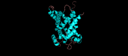
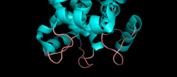
 Generic protein structure example |
Tricarboxylate transport protein, mitochondrial, also known as tricarboxylate carrier protein and citrate transport protein (CTP), is a protein that in humans is encoded by the SLC25A1 gene.[1][2][3][4] SLC25A1 belongs to the mitochondrial carrier gene family SLC25.[5][6][7] High levels of the tricarboxylate transport protein are found in the liver, pancreas and kidney. Lower or no levels are present in the brain, heart, skeletal muscle, placenta and lung.[5][7]
The tricarboxylate transport protein is located within the inner mitochondria membrane. It provides a link between the mitochondrial matrix and cytosol by transporting citrate through the impermeable inner mitochondrial membrane in exchange for malate from the cytosol.[5][6][7][8] The citrate transported out of the mitochondrial matrix by the tricarboxylate transport protein is catalyzed by citrate lyase to acetyl CoA, the starting material for fatty acid biosynthesis, and oxaloacetate.[6] As well, cytosolic NADPH + H+ necessary for fatty acid biosynthesis is generated in the reduction of oxaloacetate to malate and pyruvate by malate dehydrogenase and the malic enzyme.[7][9][10] For these reasons, the tricarboxylate transport protein is considered to play a key role in fatty acid synthesis.[6]
Structure
The structure of the tricarboxylate transport protein is consistent with the structures of other mitochondrial carriers.[5][6][8] In particular, the tricarboxylate transport protein has a tripartite structure consisting of three repeated domains that are approximately 100 amino acids in length.[5][8] Each repeat forms a transmembrane domain consisting of two hydrophobic α-helices.[5][6][11] The amino and carboxy termini are located on the cytosolic side of the inner mitochondrial membrane.[5][6] Each domain is linked by two hydrophilic loops located on the cytosolic side of the membrane.[5][6][11][12] The two α-helices of each repeated domain are connected by hydrophilic loops located on the matrix side of the membrane.[5][6][12] A salt bridge network is present on both the matrix side and cytoplasmic side of the tricarboxylate transport protein.[12]
Transport mechanism
The tricarboxylate transport protein exists in two states: a cytoplasmic state where it accepts malate from the cytoplasm and a matrix state where it accepts citrate from the mitochondrial matrix.[13] A single binding site is present near the center of the cavity of the tricarboxylate transport protein, which can be either exposed to the cytosol or the mitochondrial matrix depending on the state.[11][12][13] A substrate induced conformational change occurs when citrate enters from the matrix side and binds to the central cavity of the tricarboxylate transport protein.[5] This conformational change opens a gate on the cytosolic side and closes the gate on the matrix side.[5] Likewise, when malate enters from the cytosolic side, the matrix gate opens and the cytosolic gate closes.[5] Each side of the transporter is open and closed by the disruption and formation of the salt bridge networks, which allows access to the single binding site.[11][12][13][14][15]
Disease relevance
Mutations in this gene have been associated with the inborn error of metabolism combined D-2- and L-2-hydroxyglutaric aciduria,[16] which was the first reported case of a pathogenic mutation of the SLC25A1 gene.[12][17] Patients with D-2/L-2-hydroxyglutaric aciduria display neonatal onset metabolic encephalopathy, infantile epilepsy, global developmental delay, muscular hypotonia and early death.[12][17][18] It is believed low levels of citrate in the cytosol and high levels of citrate in the mitochondria caused by the impaired citrate transport plays a role in the disease.[12][18] In addition, increased expression of the tricarboxylate transport protein has been linked to cancer[7][19][20] and the production of inflammatory mediators.[21][22][23] Therefore, it has been suggested that inhibition of the tricarboxylate transport protein may have a therapeutic effect in chronic inflammation diseases and cancer.[22]
See also
- SLC25A1+protein,+human at the US National Library of Medicine Medical Subject Headings (MeSH)
References
- ↑ "Localization of the human mitochondrial citrate transporter protein gene to chromosome 22Q11 in the DiGeorge syndrome critical region". Genomics 29 (2): 451–6. September 1995. doi:10.1006/geno.1995.9982. PMID 8666394.
- ↑ "Organization and sequence of the human gene for the mitochondrial citrate transport protein". DNA Sequence 7 (3–4): 127–39. September 1997. doi:10.3109/10425179709034029. PMID 9254007.
- ↑ "Mitochondrial tricarboxylate and dicarboxylate-tricarboxylate carriers: from animals to plants". IUBMB Life 66 (7): 462–71. September 1997. doi:10.1002/iub.1290. PMID 25045044.
- ↑ "Entrez Gene: SLC25A1 solute carrier family 25 (mitochondrial carrier; citrate transporter), member 1". https://www.ncbi.nlm.nih.gov/sites/entrez?Db=gene&Cmd=ShowDetailView&TermToSearch=6576.
- ↑ 5.00 5.01 5.02 5.03 5.04 5.05 5.06 5.07 5.08 5.09 5.10 5.11 "The mitochondrial transporter family SLC25: identification, properties and physiopathology". Molecular Aspects of Medicine 34 (2–3): 465–84. April 2013. doi:10.1016/j.mam.2012.05.005. PMID 23266187.
- ↑ 6.0 6.1 6.2 6.3 6.4 6.5 6.6 6.7 6.8 "The mitochondrial transporter family (SLC25): physiological and pathological implications". Pflügers Archiv 447 (5): 689–709. February 2004. doi:10.1007/s00424-003-1099-7. PMID 14598172.
- ↑ 7.0 7.1 7.2 7.3 7.4 "Transcriptional Regulation of the Mitochondrial Citrate and Carnitine/Acylcarnitine Transporters: Two Genes Involved in Fatty Acid Biosynthesis and β-oxidation". Biology 2 (1): 284–303. January 2013. doi:10.3390/biology2010284. PMID 24832661.
- ↑ 8.0 8.1 8.2 Berg, Jeremy M.; Tymoczko, John L.; Gatto, Gregory J.; Stryer, Lubert (2015). Biochemistry. New York: W.H. Freeman & Company. pp. 551. ISBN 978-1-4641-2610-9.
- ↑ Voet, Donald; Voet, Judith G.; Pratt, Charlotte W. (2016). Fundamentals of Biochemistry. U.S.A.: Wiley. pp. 687–688. ISBN 978-1-118-91840-1.
- ↑ Nelson, David L.; Cox, Michael M. (2017). Principles of Biochemistry. New York: W.H. Freeman & Company. pp. 818–819. ISBN 978-1-4641-2611-6.
- ↑ 11.0 11.1 11.2 11.3 "Formation of a cytoplasmic salt bridge network in the matrix state is a fundamental step in the transport mechanism of the mitochondrial ADP/ATP carrier". Biochimica et Biophysica Acta (BBA) - Bioenergetics 1857 (1): 14–22. January 2016. doi:10.1016/j.bbabio.2015.09.013. PMID 26453935.
- ↑ 12.0 12.1 12.2 12.3 12.4 12.5 12.6 12.7 "Pathogenic mutations of the human mitochondrial citrate carrier SLC25A1 lead to impaired citrate export required for lipid, dolichol, ubiquinone and sterol synthesis". Biochimica et Biophysica Acta (BBA) - Bioenergetics 1859 (1): 1–7. January 2018. doi:10.1016/j.bbabio.2017.10.002. PMID 29031613.
- ↑ 13.0 13.1 13.2 "Mitochondrial carriers in the cytoplasmic state have a common substrate binding site". Proceedings of the National Academy of Sciences of the United States of America 103 (8): 2617–22. February 2006. doi:10.1073/pnas.0509994103. PMID 16469842. Bibcode: 2006PNAS..103.2617R.
- ↑ "The mechanism of transport by mitochondrial carriers based on analysis of symmetry". Proceedings of the National Academy of Sciences of the United States of America 105 (46): 17766–71. November 2008. doi:10.1073/pnas.0809580105. PMID 19001266. Bibcode: 2008PNAS..10517766R.
- ↑ "The conserved substrate binding site of mitochondrial carriers". Biochimica et Biophysica Acta (BBA) - Bioenergetics 1757 (9–10): 1237–48. September 2006. doi:10.1016/j.bbabio.2006.03.021. PMID 16759636.
- ↑ "Deficiency in SLC25A1, encoding the mitochondrial citrate carrier, causes combined D-2- and L-2-hydroxyglutaric aciduria". American Journal of Human Genetics 92 (4): 627–31. April 2013. doi:10.1016/j.ajhg.2013.03.009. PMID 23561848.
- ↑ 17.0 17.1 "Cerebral Organic Acid Disorders and Other Disorders of Lysine Catabolism". Inborn Metabolic Diseases. Germany: Springer. 2016. pp. 344. ISBN 978-3-662-49771-5.
- ↑ 18.0 18.1 "A novel homozygous SLC25A1 mutation with impaired mitochondrial complex V: Possible phenotypic expansion". American Journal of Medical Genetics. Part A 176 (2): 330–336. February 2018. doi:10.1002/ajmg.a.38574. PMID 29226520.
- ↑ "Quantitative metabolic flux analysis reveals an unconventional pathway of fatty acid synthesis in cancer cells deficient for the mitochondrial citrate transport protein". Metabolic Engineering 43 (Pt B): 198–207. September 2017. doi:10.1016/j.ymben.2016.11.004. PMID 27856334.
- ↑ Wan-angkan, P. (2018). "Combination of Mitochondrial and Plasma Membrane Citrate Transporter Inhibitors Inhibits De Novo Lipogenesis Pathway and Triggers Apoptosis in Hepatocellular Carcinoma Cells". BioMed Research International 2018: 3683026. doi:10.1155/2018/3683026. PMID 29546056.
- ↑ "The mitochondrial citrate carrier: a new player in inflammation". The Biochemical Journal 438 (3): 433–6. September 2011. doi:10.1042/BJ20111275. PMID 21787310. https://hal.archives-ouvertes.fr/hal-00617325.
- ↑ 22.0 22.1 "A key role of the mitochondrial citrate carrier (SLC25A1) in TNFα- and IFNγ-triggered inflammation". Biochimica et Biophysica Acta (BBA) - Gene Regulatory Mechanisms 1839 (11): 1217–1225. November 2014. doi:10.1016/j.bbagrm.2014.07.013. PMID 25072865.
- ↑ "Acetylation of human mitochondrial citrate carrier modulates mitochondrial citrate/malate exchange activity to sustain NADPH production during macrophage activation". Biochimica et Biophysica Acta (BBA) - Bioenergetics 1847 (8): 729–38. August 2015. doi:10.1016/j.bbabio.2015.04.009. PMID 25917893.
Further reading
- "Large-scale mapping of human protein-protein interactions by mass spectrometry". Molecular Systems Biology 3 (1): 89. 2007. doi:10.1038/msb4100134. PMID 17353931.
- "Towards a proteome-scale map of the human protein-protein interaction network". Nature 437 (7062): 1173–8. October 2005. doi:10.1038/nature04209. PMID 16189514. Bibcode: 2005Natur.437.1173R.
- "A transcription map of the DiGeorge and velo-cardio-facial syndrome minimal critical region on 22q11". Human Molecular Genetics 5 (6): 789–800. June 1996. doi:10.1093/hmg/5.6.789. PMID 8776594.
- "Cloning, genomic organization, and chromosomal localization of human citrate transport protein to the DiGeorge/velocardiofacial syndrome minimal critical region". Genomics 33 (2): 271–6. April 1996. doi:10.1006/geno.1996.0191. PMID 8660975.
- "Mechanisms of divergent effects of activated peroxisome proliferator-activated receptor-γ on mitochondrial citrate carrier expression in 3T3-L1 fibroblasts and mature adipocytes". Biochimica et Biophysica Acta (BBA) - Molecular and Cell Biology of Lipids 1831 (6): 1027–36. June 2013. doi:10.1016/j.bbalip.2013.01.014. PMID 23370576.
This article incorporates text from the United States National Library of Medicine, which is in the public domain.
 |


