Biology:Beta-glucuronidase
| β-Glucuronidase | |||||||||
|---|---|---|---|---|---|---|---|---|---|
 | |||||||||
| Identifiers | |||||||||
| EC number | 3.2.1.31 | ||||||||
| CAS number | 9001-45-0 | ||||||||
| Databases | |||||||||
| IntEnz | IntEnz view | ||||||||
| BRENDA | BRENDA entry | ||||||||
| ExPASy | NiceZyme view | ||||||||
| KEGG | KEGG entry | ||||||||
| MetaCyc | metabolic pathway | ||||||||
| PRIAM | profile | ||||||||
| PDB structures | RCSB PDB PDBe PDBsum | ||||||||
| Gene Ontology | AmiGO / QuickGO | ||||||||
| |||||||||
| β-Glucuronidase | |
|---|---|
 β-Glucuronidase asymmetric unit showing active site residues Glu451, Tyr504, and Glu540, along with the potentially supporting Asn450 residue[1] | |
| Identifiers | |
| Symbol | GUSB |
| NCBI gene | 2990 |
| HGNC | 4696 |
| OMIM | 611499 |
| RefSeq | NM_000181 |
| UniProt | P08236 |
| Other data | |
| EC number | 3.2.1.31 |
| Locus | Chr. 7 q11.21 |
β-Glucuronidases are members of the glycosidase family of enzymes that catalyze breakdown of complex carbohydrates.[2] Human β-glucuronidase is a type of glucuronidase (a member of glycosidase Family 2) that catalyzes hydrolysis of β-D-glucuronic acid residues from the non-reducing end of mucopolysaccharides (also referred to as glycosaminoglycans) such as heparan sulfate.[2][3][4] Human β-glucuronidase is located in the lysosome.[5] In the gut, brush border β-glucuronidase converts conjugated bilirubin to the unconjugated form for reabsorption. β-Glucuronidase is also present in breast milk, which contributes to neonatal jaundice. The protein is encoded by the GUSB gene in humans[6][7] and by the uidA gene in bacteria.[8]
Structure
Human β-glucuronidase is synthesized as an 80 kDa monomer (653 amino acids) before proteolysis removes 18 amino acids from the C-terminal end to form a 78 kDa monomer.[9][10] β-Glucuronidase exists as a 332 kDa homotetramer.[11] β-Glucuronidase contains several notable structural formations, including a type of β-barrel known as a jelly roll barrel and a TIM barrel.[1]
Mechanism of catalysis
Human β-glucuronidase is homologous to the Escherichia coli enzyme β-galactosidase.[12][13] This homologous relationship, along with the knowledge that glycosidases often perform hydrolysis catalyzed by two acidic residues, enabled the development of a mechanistic hypothesis. This hypothesis proposes that the two glutamic acid residues Glu540 and Glu451 are the nucleophilic and acidic residues, respectively, and that the tyrosine residue Tyr504 is also involved in catalysis. In support of this hypothesis, experimental mutations in any of these three residues result in large decreases of enzymatic activity. Increased activity of an E451A mutant enzyme (where Glu451 is replaced with an alanine residue) after addition of azide is consistent with Glu451 as the acid/base residue.[14] Using analysis of labeled β-glucuronidase peptides after hydrolysis of a substrate that enters a very stable intermediate stage, researchers have determined that Glu540 is the nucleophilic residue.[15]
Though the particular type of nucleophilic substitution employed by β-glucuronidase is unclear, evidence for the mechanisms of their homologues in the glycosidase family suggests that these reactions are qualitatively SN2 reactions. The reactions proceed through a transition state with oxocarbenium ion characteristics. Initially, these mechanisms, because of this oxocarbenium characteristic of the transition state, were suggested to be SN1 reactions proceeding through a discrete oxocarbenium ion intermediate. However, more recent evidence suggests that these oxocarbenium ion states have lifetimes of 10 femtoseconds - 0.1 nanoseconds (similar to that of a bond vibration period). These lifetimes are too short to assign to a reaction intermediate. From this evidence, it appears that these reactions, while having an SN1 appearance due to the oxocarbenium ion characteristics of their transition states, must be qualitatively SN2 reactions.[2]
The specific activity of Tyr504 in the catalytic mechanism is unclear.[14] Through comparison to the structural data of the homologous enzyme xylanase, it has been suggested that Tyr504 of β-glucuronidase might stabilize the leaving nucleophile (Glu540) or modulate its activity.[16]
In addition to these residues, a conserved asparagine residue (Asn450) has been suggested to stabilize the substrate through the action of a hydrogen bond at the 2-hydroxyl group of the sugar substrate.[11][17]
-
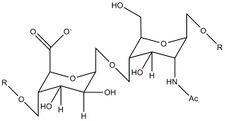 Repeating unit of the heparan sulfate substrate of β-glucuronidase
 -
![Surface depiction of active site pocket of β-glucuronidase with catalytic residues shown[1]](/wiki/images/thumb/9/98/Beta_Glucuronidase_Active_Site_Pocket.jpg/225px-Beta_Glucuronidase_Active_Site_Pocket.jpg) Surface depiction of active site pocket of β-glucuronidase with catalytic residues shown[1]
Surface depiction of active site pocket of β-glucuronidase with catalytic residues shown[1] -
![Mechanism of β-glucuronidase hydrolysis of a sugar substrate with high energy transition states showing oxocarbenium ion character depicted[15]](/wiki/images/thumb/7/76/Mechanism_Of_Beta_Glucuronidase.JPG/225px-Mechanism_Of_Beta_Glucuronidase.JPG) Mechanism of β-glucuronidase hydrolysis of a sugar substrate with high energy transition states showing oxocarbenium ion character depicted[15]
Mechanism of β-glucuronidase hydrolysis of a sugar substrate with high energy transition states showing oxocarbenium ion character depicted[15] -
![Potential stabilization of the nucleophilic residue Glu540 by Tyr504 in β-glucuronidase[16]](/wiki/images/thumb/4/4c/File-Predicted_Tyr_504_activity_in_Beta_Glucuronidase.JPG/225px-File-Predicted_Tyr_504_activity_in_Beta_Glucuronidase.JPG) Potential stabilization of the nucleophilic residue Glu540 by Tyr504 in β-glucuronidase[16]
Potential stabilization of the nucleophilic residue Glu540 by Tyr504 in β-glucuronidase[16] -
![Predicted activity of the conserved Asn450 residue in stabilization of the β-glucuronidase sugar substrate[11][17]](/wiki/images/thumb/1/1e/Predicted_Asn_450_activity_in_beta_glucuronidase.JPG/175px-Predicted_Asn_450_activity_in_beta_glucuronidase.JPG)
-
![Potential salt bridge between Glu352 and Arg216 in human beta-glucuronidase[1][18]](/wiki/images/thumb/8/8e/Possible_Salt_Bridge_Human_Beta_Glucuronidase.jpg/225px-Possible_Salt_Bridge_Human_Beta_Glucuronidase.jpg)

![Surface depiction of active site pocket of β-glucuronidase with catalytic residues shown[1]](/wiki/File:Beta_Glucuronidase_Active_Site_Pocket.jpg)
![Mechanism of β-glucuronidase hydrolysis of a sugar substrate with high energy transition states showing oxocarbenium ion character depicted[15]](/wiki/File:Mechanism_Of_Beta_Glucuronidase.JPG)
![Potential stabilization of the nucleophilic residue Glu540 by Tyr504 in β-glucuronidase[16]](/wiki/File:File-Predicted_Tyr_504_activity_in_Beta_Glucuronidase.JPG)
![Predicted activity of the conserved Asn450 residue in stabilization of the β-glucuronidase sugar substrate[11][17]](/wiki/File:Predicted_Asn_450_activity_in_beta_glucuronidase.JPG)
![Potential salt bridge between Glu352 and Arg216 in human beta-glucuronidase[1][18]](/wiki/File:Possible_Salt_Bridge_Human_Beta_Glucuronidase.jpg)
Sly syndrome
Deficiencies in β-glucuronidase result in the autosomal recessive inherited metabolic disease known as Sly syndrome or Mucopolysaccharidosis VII. A deficiency in this enzyme results in the build-up of non-hydrolyzed mucopolysaccharides in the patient. This disease can be extremely debilitating for the patient or can result in hydrops fetalis prior to birth. In addition, mental retardation, short stature, coarse facial features, spinal abnormalities, and enlargement of liver and spleen are observed in surviving patients.[5] This disease has been modeled in a strain of mice as well as a family of dogs.[19][20] More recently researchers have discovered a feline family that exhibits deficiencies in β-glucuronidase activity. The source of this reduction of activity has been identified as an E351K mutation (Glu351 is mutated to a lysine residue). Glu351 is conserved in mammalian species, which suggests an important function for this residue. Examination of the human X-ray crystal structure suggests that this residue (Glu352 in the human enzyme), which is buried deep within the TIM barrel domain, may be important for stabilization of the tertiary structure of the enzyme.[18] In the crystal structure, it appears that Arg216, a member of the jelly roll domain of the protein, forms a salt bridge with Glu352; therefore, Glu352 is likely involved in stabilizing the interaction between two different three-dimensional domains of the enzyme.[1]
Use as a reporter gene
In molecular biology, β-glucuronidase is used as a reporter gene to monitor gene expression in mammalian and plant cells. Monitoring β-glucuronidase activity through the use of a GUS assay allows determination of the spatial and temporal expression of the gene in question.[21]
- Molecular graphics images were produced using the Chimera package from the Resource for Biocomputing, Visualization, and Informatics at the University of California, San Francisco. [22]
See also
- Alpha-glucuronidase
- Glucuronosyl-disulfoglucosamine glucuronidase
- Glycyrrhizinate beta-glucuronidase
References
- ↑ 1.0 1.1 1.2 1.3 1.4 1.5 PDB: 1BHG; "Structure of human β-glucuronidase reveals candidate lysosomal targeting and active-site motifs". Nature Structural Biology 3 (4): 375–81. April 1996. doi:10.1038/nsb0496-375. PMID 8599764.
- ↑ 2.0 2.1 2.2 Comprehensive Biological Catalysis. 1. Manchester, UK: Academic Press. 1998. pp. 119–138. ISBN 978-0-12-646864-9. https://archive.org/details/encyclopediaofhu0003unse/page/119.
- ↑ "Mechanisms of enzymatic glycoside hydrolysis". Current Opinion in Structural Biology 4 (6): 885–92. December 1994. doi:10.1016/0959-440X(94)90271-2. PMID 7712292.
- ↑ Sinnott ML (1990). "Catalytic mechanisms of enzymic glycosyl transfer". Chem Rev 90 (7): 1171–1202. doi:10.1021/cr00105a006.
- ↑ 5.0 5.1 Nyhan, William L.; Barshop, Bruce; Ozand, Pinar (2005). Atlas of Metabolic Diseases (2 ed.). London, UK: Hodder Arnold. pp. 501–503, 546–550. ISBN 978-0-340-80970-9.
- ↑ "Cloning, sequencing, and expression of cDNA for human β-glucuronidase". Proceedings of the National Academy of Sciences of the United States of America 84 (3): 685–9. February 1987. doi:10.1073/pnas.84.3.685. PMID 3468507. Bibcode: 1987PNAS...84..685O.
- ↑ "Entrez Gene: GUSB glucuronidase, beta". https://www.ncbi.nlm.nih.gov/sites/entrez?Db=gene&Cmd=ShowDetailView&TermToSearch=2990.
- ↑ "Distribution of uidA gene sequences in Escherichia coli isolates in water sources and comparison with the expression of beta-glucuronidase activity in 4-methylumbelliferyl-β-D-glucuronide media". Applied and Environmental Microbiology 59 (7): 2271–6. July 1993. doi:10.1128/AEM.59.7.2271-2276.1993. PMID 8357258. Bibcode: 1993ApEnM..59.2271M.
- ↑ "C-terminal processing of human beta-glucuronidase. The propeptide is required for full expression of catalytic activity, intracellular retention, and proper phosphorylation". The Journal of Biological Chemistry 268 (30): 22627–33. October 1993. doi:10.1016/S0021-9258(18)41574-8. PMID 8226771.
- ↑ "The role of glycosylation and phosphorylation in the expression of active human β-glucuronidase". The Journal of Biological Chemistry 268 (16): 12193–8. June 1993. doi:10.1016/S0021-9258(19)50325-8. PMID 8505339.
- ↑ 11.0 11.1 11.2 "Crystallization and preliminary X-ray analysis of endoglucanase from Pyrococcus horikoshii". Acta Crystallographica. Section F, Structural Biology and Crystallization Communications 64 (Pt 12): 1169–71. December 2008. doi:10.1107/S1744309108036919. PMID 19052378.
- ↑ "New families in the classification of glycosyl hydrolases based on amino acid sequence similarities". The Biochemical Journal 293 ( Pt 3) (3): 781–8. August 1993. doi:10.1042/bj2930781. PMID 8352747.
- ↑ "A classification of glycosyl hydrolases based on amino acid sequence similarities". The Biochemical Journal 280 ( Pt 2) (2): 309–16. December 1991. doi:10.1042/bj2800309. PMID 1747104.
- ↑ 14.0 14.1 "Active site residues of human β-glucuronidase. Evidence for Glu(540) as the nucleophile and Glu(451) as the acid-base residue". The Journal of Biological Chemistry 274 (33): 23451–5. August 1999. doi:10.1074/jbc.274.33.23451. PMID 10438523.
- ↑ 15.0 15.1 "Identification of Glu-540 as the catalytic nucleophile of human beta-glucuronidase using electrospray mass spectrometry". The Journal of Biological Chemistry 273 (51): 34057–62. December 1998. doi:10.1074/jbc.273.51.34057. PMID 9852062.
- ↑ 16.0 16.1 "EzCatDB: T00066". EzCatDB: A Database of Catalytic Mechanisms. http://mbs.cbrc.jp/EzCatDB/search/get.do?dbcode=T00066.
- ↑ 17.0 17.1 "Conserved catalytic machinery and the prediction of a common fold for several families of glycosyl hydrolases". Proceedings of the National Academy of Sciences of the United States of America 92 (15): 7090–4. July 1995. doi:10.1073/pnas.92.15.7090. PMID 7624375. Bibcode: 1995PNAS...92.7090H.
- ↑ 18.0 18.1 "Molecular basis of feline beta-glucuronidase deficiency: an animal model of mucopolysaccharidosis VII". Genomics 58 (2): 121–8. June 1999. doi:10.1006/geno.1999.5825. PMID 10366443.
- ↑ "Murine mucopolysaccharidosis type VII. Characterization of a mouse with beta-glucuronidase deficiency". The Journal of Clinical Investigation 83 (4): 1258–66. April 1989. doi:10.1172/JCI114010. PMID 2495302.
- ↑ "Beta-glucuronidase deficiency in a dog: a model of human mucopolysaccharidosis VII". Pediatric Research 18 (10): 980–4. October 1984. doi:10.1203/00006450-198410000-00014. PMID 6436780.
- ↑ "Vectors with the gus reporter gene for identifying and quantitating promoter regions in Saccharomyces cerevisiae". Gene 154 (1): 105–7. February 1995. doi:10.1016/0378-1119(94)00845-J. PMID 7867935.
- ↑ "UCSF Chimera--a visualization system for exploratory research and analysis". Journal of Computational Chemistry 25 (13): 1605–12. October 2004. doi:10.1002/jcc.20084. PMID 15264254. http://www.cgl.ucsf.edu/home/tef/pubs/chimera.pdf.
Further reading
- "Elevated serum beta-glucuronidase reflects hepatic lysosomal fragility following toxic liver injury in rats". Biochemistry and Cell Biology 86 (3): 235–43. June 2008. doi:10.1139/O08-038. PMID 18523484.
- "Human beta-glucuronidase deficiency mucopolysaccharidosis: identification of cross-reactive antigen in cultured fibroblasts of deficient patients by enzyme immunoassay". The Journal of Clinical Investigation 59 (1): 97–105. January 1977. doi:10.1172/JCI108627. PMID 401508.
- "Characterization of the subunits and sugar moiety of human placental and leukemic beta-glucuronidase". Biological Chemistry Hoppe-Seyler 373 (1): 57–62. January 1992. doi:10.1515/bchm3.1992.373.1.57. PMID 1311180.
- "Reversal of pathology in murine mucopolysaccharidosis type VII by somatic cell gene transfer". Nature 360 (6406): 749–53. 1993. doi:10.1038/360749a0. PMID 1465145.
- "Mucopolysaccharidosis type VII: characterization of mutations and molecular heterogeneity". American Journal of Human Genetics 48 (1): 89–96. January 1991. PMID 1702266.
- "Analysis of the 5' flanking region of the human beta-glucuronidase gene". Genomics 10 (4): 1009–18. August 1991. doi:10.1016/0888-7543(91)90192-H. PMID 1916806.
- "Phosphorylation of beta-glucuronidases from human normal liver and hepatoma by cAMP-dependent protein kinase". The Journal of Biological Chemistry 263 (12): 5884–9. April 1988. doi:10.1016/S0021-9258(18)60648-9. PMID 2833520.
- "Isolation and expression in Escherichia coli of a cDNA clone encoding human beta-glucuronidase". Gene 34 (1): 105–10. 1985. doi:10.1016/0378-1119(85)90300-2. PMID 3924735.
- "Human hepatic beta-glucuronidase: an enzyme kinetic study". Enzyme 33 (1): 9–17. 1985. doi:10.1159/000469398. PMID 3987656.
- "Mutational analysis of a patient with mucopolysaccharidosis type VII, and identification of pseudogenes". American Journal of Human Genetics 52 (3): 517–26. March 1993. PMID 7680524.
- "Molecular analysis of a patient with hydrops fetalis caused by beta-glucuronidase deficiency, and evidence for additional pseudogenes". Human Mutation 2 (6): 443–5. 1994. doi:10.1002/humu.1380020604. PMID 8111412.
- "Mutational studies in a patient with the hydrops fetalis form of mucopolysaccharidosis type VII". Human Mutation 2 (6): 446–57. 1994. doi:10.1002/humu.1380020605. PMID 8111413.
- "Oligo-capping: a simple method to replace the cap structure of eukaryotic mRNAs with oligoribonucleotides". Gene 138 (1–2): 171–4. January 1994. doi:10.1016/0378-1119(94)90802-8. PMID 8125298.
- "Correction of lysosomal storage in the liver and spleen of MPS VII mice by implantation of genetically modified skin fibroblasts". Nature Genetics 4 (2): 154–9. June 1993. doi:10.1038/ng0693-154. PMID 8348154.
- "The role of glycosylation and phosphorylation in the expression of active human beta-glucuronidase". The Journal of Biological Chemistry 268 (16): 12193–8. June 1993. doi:10.1016/S0021-9258(19)50325-8. PMID 8505339.
- "Biochemical characterization of liver microsomal, Golgi, lysosomal, and serum beta-glucuronidases in dibutyl phosphate-treated rats". Journal of Biochemistry 118 (1): 56–66. July 1995. doi:10.1093/oxfordjournals.jbchem.a124892. PMID 8537326.
- "Structure of human beta-glucuronidase reveals candidate lysosomal targeting and active-site motifs". Nature Structural Biology 3 (4): 375–81. April 1996. doi:10.1038/nsb0496-375. PMID 8599764.
- "Molecular analysis of patients with beta-glucuronidase deficiency presenting as hydrops fetalis or as early mucopolysaccharidosis VII". American Journal of Human Genetics 58 (3): 457–71. March 1996. PMID 8644704.
- "Normalization and subtraction: two approaches to facilitate gene discovery". Genome Research 6 (9): 791–806. September 1996. doi:10.1101/gr.6.9.791. PMID 8889548.
- "Subtle differences between human and rabbit neutrophil receptors shown by the secretagogue activity of constrained formyl peptides". Archives of Biochemistry and Biophysics 337 (2): 267–74. January 1997. doi:10.1006/abbi.1996.9791. PMID 9016822.
PDB gallery | |
|---|---|
|

External links
- Glucuronidase at the US National Library of Medicine Medical Subject Headings (MeSH)
- Updated research on reporter glucuronidase and other reporters from Reportergene
- Database of Catalytic Mechanism Research and other information on beta-glucuronidase
 |
