Biology:Cancer cell
Cancer cells are cells that divide continually, forming solid tumors or flooding the blood or lymph with abnormal cells. Cell division is a normal process used by the body for growth and repair. A parent cell divides to form two daughter cells, and these daughter cells are used to build new tissue or to replace cells that have died because of aging or damage. Healthy cells stop dividing when there is no longer a need for more daughter cells, but cancer cells continue to produce copies. They are also able to spread from one part of the body to another in a process known as metastasis.[1]

Classification
There are different categories of cancer cell, defined according to the cell type from which they originate.[2]
- Carcinoma, the majority of cancer cells are epithelial in origin, beginning in a tissue that lines the inner or outer surfaces of the body.
- Leukaemia, originate in the tissues responsible for producing new blood cells, most commonly in the bone marrow.
- Lymphoma and myeloma, derived from cells of the immune system.
- Sarcoma, originating in connective tissue, including fat, muscle and bone.
- Central nervous system, derived from cells of the brain and spinal cord.
- Mesothelioma, originating in the mesothelium; the lining of body cavities.
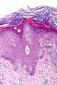
Carcinoma
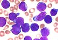
Leukaemia
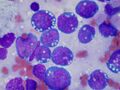
Lymphoma
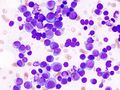
Myeloma
Sarcoma
Mesothelioma


_MG_stain.jpg)


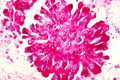
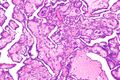
Histology

Cancer cells have distinguishing histological features visible under the microscope. The nucleus is often large and irregular, and the cytoplasm may also display abnormalities.[3]
Nucleus
The shape, size, protein composition, and texture of the nucleus are often altered in malignant cells. The nucleus may acquire grooves, folds or indentations, chromatin may aggregate or disperse, and the nucleolus can become enlarged. In normal cells, the nucleus is often round or solid in shape, but in cancer cells the outline is often irregular. Different combinations of abnormalities are characteristic of different cancer types, to the extent that nuclear appearance can be used as a marker in cancer diagnostics and staging.[4]
Causes
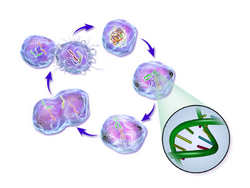
Life cycle of a cancer cell

- Cancer cells are created when the genes responsible for regulating cell division are damaged. Carcinogenesis is caused by mutation and epimutation of the genetic material of normal cells, which upsets the normal balance between proliferation and cell death. This results in uncontrolled cell division in the body. The uncontrolled and often rapid proliferation of cells can lead to benign or malignant tumours (cancer). Benign tumors do not spread to other parts of the body or invade other tissues. Malignant tumors can invade other organs, spread to distant locations (metastasis) and become life-threatening.
More than one mutation is necessary for carcinogenesis. In fact, a series of several mutations to certain classes of genes is usually required before a normal cell will transform into a cancer cell.[5]
Damage to DNA can be caused by exposure to radiation, chemicals, and other environmental sources, but mutations also accumulate naturally over time through uncorrected errors in DNA transcription, making age another risk factor. Oncoviruses can cause certain types of cancer, and genetics are also known to play a role.[6]
Stem cell research suggests that excess SP2 protein may turn stem cells into cancer cells.[7] However, a lack of particular co-stimulated molecules that aid in the way antigens react with lymphocytes can impair the natural killer cells' function, ultimately leading to cancer.[8][failed verification]
DNA repair and mutation
When a cell is deficient in the capacity to repair DNA damages, such damages tend to be retained within the cell at an increased level. These damages, upon replication of the cell’s DNA, may cause replication errors, including mutations that lead to cancer. Numerous inherited DNA repair disorders have been described that increase cancer risk (see Wikipedia article DNA repair-deficiency disorder). In addition, particular DNA repair enzymes have been found to be deficient in multiple cancers. For example, deficient expression of the DNA repair enzyme O-6-methylguanine-DNA methyltransferase is observed in several different kinds of cancer (see Wikipedia article O-6-methylguanine-DNA methyltransferase). Although a DNA repair deficiency can predispose a cell lineage to develop cancer, increased (rather than decreased) expression of a repair capability may also emerge in the progression of cancer cell lineages, and this capability may be clinically important as reviewed by Lingg et al.[9] For instance, the DNA repair gene DMC1 encodes a protein that is normally expressed only in cells undergoing meiosis where it helps maintain an undamaged germ-line. However, DMC1 is also expressed in various cancer cell lines including cervical, breast, and lymphoma cancer cell lines.[9] Expression of meiotic DNA repair genes such as DMC1 may promote tumor cell growth by dealing with endogenous DNA damage within the tumor, and may also diminish the effectiveness of anticancer therapy, such as radiation therapy.[9]
Pathology
Cells playing roles in the immune system, such as T-cells, are thought to use a dual receptor system when they determine whether or not to kill sick or damaged human cells. If a cell is under stress, turning into tumors, or infected, molecules including MIC-A and MIC-B are produced so that they can attach to the surface of the cell.[8] These work to help macrophages detect and kill cancer cells.[10]
Discovery
Early evidence of human cancer can be interpreted from Egyptian papers (1538 BCE) and mummified remains.[11] In 2016, a 1.7 million year old osteosarcoma was reported by Edward John Odes (a doctoral student in Anatomical Sciences from Witwatersrand Medical School, South Africa) and colleagues, representing the oldest documented malignant hominin cancer.[12]
The understanding of cancer was significantly advanced during the Renaissance period and in to the Age of Discovery. Sir Rudolf Virchow, a German biologist and politician, studied microscopic pathology, and linked his observations to illness. He is described as "the founder of cellular pathology".[13] In 1845, Virchow and John Hughes Bennett independently observed abnormal increase in white blood cells in patients. Virchow correctly identified the condition as blood disease, and named it leukämie in 1847 (later anglicised to leukemia).[14][15][16] In 1857, he was the first to describe a type of tumour called chordoma that originated from the clivus (at the base of the skull).[17][18]
Telomerase
Cancer cells have unique features that make them "immortal" according to some researchers. The enzyme telomerase is used to extend the cancer cell's life span. While the telomeres of most cells shorten after each division, eventually causing the cell to die, telomerase extends the cell's telomeres. This is a major reason that cancer cells can accumulate over time, creating tumors.
Cancer stem cells and drug resistance

Scientists have discovered a molecule on the surface of tumors that appears to promote drug resistance—by converting the tumor cells back into a stem cell-like state.
When the tumor cells began to exhibit drug resistance, the cells were simultaneously transforming into a stem cell-like state, which made them impervious to the drugs. It appeared that the treatment itself was driving this transformation by activating a specific molecular pathway. Luckily, several existing drugs, such as Bortezomib for example, can attack this pathway and reverse the cellular transformation, thus 're-sensitizing' the tumor to treatment.[19][20][21]
Treatment
In February 2019, medical scientists announced that iridium attached to albumin, creating a photosensitized molecule, can penetrate cancer cells and, after being irradiated with light (a process called photodynamic therapy), destroy the cancer cells.[22][23]
See also
References
- ↑ "National Cancer Institute: is this cancer?". 2007-09-17. http://www.cancer.gov/about-cancer/understanding/what-is-cancer.
- ↑ "Histological types of cancer - CRS - Cancer Research Society". http://www.crs-src.ca/page.aspx?pid=1765.
- ↑ "Comparative Oncology" (in en). Tumor Cell Morphology. The Publishing House of the Romanian Academy. 2007. https://www.ncbi.nlm.nih.gov/books/NBK9553/.
- ↑ "Nuclear structure in cancer cells". Nature Reviews. Cancer 4 (9): 677–687. September 2004. doi:10.1038/nrc1430. PMID 15343274.
- ↑ "A genetic model for colorectal tumorigenesis". Cell 61 (5): 759–767. June 1990. doi:10.1016/0092-8674(90)90186-I. PMID 2188735.
- ↑ What causes cancer? : Cancer Research UK : CancerHelp UK. Cancerhelp.org.uk (2010-07-15). Retrieved on 2010-12-01.
- ↑ Too much SP2 protein turns stem cells into 'EVIL TWIN' cancer cells. Sciencedaily.com (2010-10-27). Retrieved on 2010-12-01.
- ↑ 8.0 8.1 "The Innate Immune System: NK Cells". Community College of Baltimore County. http://student.ccbcmd.edu/courses/bio141/lecguide/unit4/innate/nkcell.html.
- ↑ 9.0 9.1 9.2 Lingg, L.; Rottenberg, S.; Francica, P. (2022). "Meiotic Genes and DNA Double Strand Break Repair in Cancer". Frontiers in Genetics 13: 831620. doi:10.3389/fgene.2022.831620. PMID 35251135.
- ↑ The Adaptive Immune System: Ways That Antibodies Help to Defend the Body - Antibody-Dependent Cellular Cytotoxicity (ADCC) . Student.ccbcmd.edu. Retrieved on 2010-12-01.
- ↑ "Cancer: an old disease, a new disease or something in between?" (in En). Nature Reviews. Cancer 10 (10): 728–733. October 2010. doi:10.1038/nrc2914. PMID 20814420.
- ↑ "Earliest hominin cancer: 1.7-million-year-old osteosarcoma from Swartkrans Cave, South Africa" (in en). South African Journal of Science 112 (7/8): 5. 2016. doi:10.17159/sajs.2016/20150471. ISSN 1996-7489. http://sajs.co.za/earliest-hominin-cancer-1-7-million-year-old-osteosarcoma-swartkrans-cave-south-africa/edward-j-odes-patrick-s-randolph-quinney-maryna-steyn-zach-throckmorton-jacqueline-s-smilg-bernhard. Retrieved 2016-08-01.
- ↑ "History of cancer". University of Bergen. 23 March 1999. http://www.uib.no/med/avd/miapr/arvid/MOD2/Arvid/Patofysiologi/history_of_cancer.pdf.
- ↑ "John Hughes Bennett, Rudolph Virchow... and Alfred Donné: the first description of leukemia". The Hematology Journal 2 (1): 1. 2001. doi:10.1038/sj/thj/6200090. PMID 11920227.
- ↑ "The discovery and early understanding of leukemia". Leukemia Research 36 (1): 6–13. January 2012. doi:10.1016/j.leukres.2011.09.028. PMID 22033191.
- ↑ The Emperor of All Maladies: A Biography of Cancer. Simon and Schuster. 16 November 2010. ISBN 978-1-4391-0795-9. https://books.google.com/books?id=5rF_31RVTnMC. Retrieved 6 September 2011.
- ↑ "Sacrococcygeal chordoma". JAMA: The Journal of the American Medical Association 80 (19): 1369. May 1923. doi:10.1001/jama.1923.02640460019007.
- ↑ "Chordoma: retrospective analysis of 24 cases". Sao Paulo Medical Journal = Revista Paulista de Medicina 114 (6): 1312–1316. 1996. doi:10.1590/S1516-31801996000600006. PMID 9269106.
- ↑ "Drug resistance and Cancer stem cells". Cell Communication and Signaling 19 (1): 19. February 2021. doi:10.1186/s12964-020-00627-5. PMID 33588867.
- ↑ "Tumor Cells Become Drug Resistant by Reverting to a Stem Cell-Like State". 23 April 2014. https://blog.cirm.ca.gov/2014/04/23/tumor-cells-become-drug-resistant-by-reverting-to-a-stem-cell-like-state-new-study-finds/.
- ↑ "An integrin β3-KRAS-RalB complex drives tumour stemness and resistance to EGFR inhibition". Nature Cell Biology 16 (5): 457–468. May 2014. doi:10.1038/ncb2953. PMID 24747441.
- ↑ University of Warwick (3 February 2019). "Simply shining light on dinosaur metal compound kills cancer cells". EurekAlert!. https://www.eurekalert.org/pub_releases/2019-02/uow-ssl013119.php.
- ↑ "Nucleus-Targeted Organoiridium-Albumin Conjugate for Photodynamic Cancer Therapy". Angewandte Chemie 58 (8): 2350–2354. February 2019. doi:10.1002/anie.201813002. PMID 30552796.
Further reading
- Schwarzer, K.; Foerster, M.; Steiner, T.; Hermann, I. M.; Straube, E. (2010). "BCG strain S4-Jena: An early BCG strain is capable to reduce the proliferation of bladder cancer cells by induction of apoptosis". Cancer Cell International 10: 21. doi:10.1186/1475-2867-10-21. PMID 20587032.
External links
- The History of Cancer . Cancer.org. Retrieved on 2010-12-01
{{Navbox
| name = Tumors
| title = Overview of tumors, cancer and oncology (C00–D48, 140–239)
| state = collapsed
| listclass = hlist
| group1 = Conditions
| list1 =
| Benign tumors | |
|---|---|
| Malignant progression | |
| Topography | |
| Histology | |
| Other |
| group2 = Staging/grading
| list2 =
| group3 = Carcinogenesis
| list3 =
- Cancer cell
- Carcinogen
- [[Biology:Tumor suppressor Tumor suppressor genes/oncogenes
- Clonally transmissible cancer
- Oncovirus
- Carcinogenic bacteria
| group4 = Misc.
| list4 =
}}
 |  |

