Medicine:Osteoporosis
Osteoporosis is a systemic skeletal disorder characterized by low bone mass (osteopenia), micro-architectural deterioration of bone tissue leading to more porous bone, and consequent increase in fracture risk. Bones undergo continuous remodeling: osteoclasts resorb old bone, and osteoblasts synthesize new bone. With advancing age, the rate of resorption exceeds that of bone formation, causing bones to lose density and become more susceptible to fractures.[1]
It is the most common reason for a broken bone among the elderly.[2] Bones that commonly break include the vertebrae in the spine, the bones of the forearm, the wrist, and the hip.[3][4]
Until a broken bone occurs, there are typically no symptoms. Bones may weaken to such a degree that a break may occur with minor stress or spontaneously. After the broken bone heals, some people may have chronic pain and a decreased ability to carry out normal activities.[2]
Osteoporosis may be due to lower-than-normal maximum bone mass and greater-than-normal bone loss. Bone loss increases after menopause in women due to lower levels of estrogen,[5] and after andropause in older men due to lower levels of testosterone.[6] Osteoporosis may also occur due to several diseases or treatments, including alcoholism, anorexia or underweight, hyperparathyroidism, hyperthyroidism, kidney disease, and after oophorectomy (surgical removal of the ovaries). Certain medications increase the rate of bone loss, including some antiseizure medications, chemotherapy, proton pump inhibitors, selective serotonin reuptake inhibitors, glucocorticosteroids, and overzealous levothyroxine suppression therapy. Smoking and sedentary lifestyle are also recognized as major risk factors.[2] Osteoporosis is defined as a bone density of 2.5 standard deviations below that of a young adult. This is typically measured by dual-energy X-ray absorptiometry (DXA or DEXA).[7]
Prevention of osteoporosis includes a proper diet during childhood, hormone replacement therapy for menopausal women, and efforts to avoid medications that increase the rate of bone loss. Efforts to prevent broken bones in those with osteoporosis include a good diet, exercise, and fall prevention. Lifestyle changes such as stopping smoking and not drinking alcohol may help.[2] Bisphosphonate medications are useful to decrease future broken bones in those with previous broken bones due to osteoporosis. In those with osteoporosis but no previous broken bones, they have been shown to be less effective.[8][needs update][9][10] They do not appear to affect the risk of death.[11]
Osteoporosis becomes more common with age. About 15% of Caucasians in their 50s and 70% of those over 80 are affected.[12] It is more common in women than men.[2] In the developed world, depending on the method of diagnosis, 2% to 8% of males and 9% to 38% of females are affected.[13] Rates of disease in the developing world are unclear.[14] About 22 million women and 5.5 million men in the European Union had osteoporosis in 2010.[15] In the United States in 2010, about 8 million women and between 1 and 2 million men had osteoporosis.[13][16] White and Asian people are at greater risk for low bone mineral density due to their lower serum vitamin D levels and less vitamin D synthesis at certain latitudes.[2] The word "osteoporosis" is from the Greek terms for "porous bones".[17]
Signs and symptoms

Osteoporosis has no symptoms, and the person usually does not know that they have osteoporosis until a bone is broken. Osteoporotic fractures occur in situations where healthy people would not normally break a bone; they are therefore regarded as fragility fractures. Typical fragility fractures occur in the vertebral column, rib, hip and wrist.[18] Examples of situations where people would not normally break a bone include a fall from standing height, normal day-to-day activities such as lifting, bending, or coughing.[18]
Fractures
Fractures are a common complication of osteoporosis and can result in disability.[19] Acute and chronic pain in the elderly is often attributed to fractures from osteoporosis and can lead to further disability and early mortality.[20] These fractures may also be asymptomatic.[21] The most common osteoporotic fractures are of the wrist, spine, shoulder, and hip. The symptoms of a vertebral collapse ("compression fracture") are sudden back pain, often with radicular pain (shooting pain due to nerve root compression) and rarely with spinal cord compression or cauda equina syndrome. Multiple vertebral fractures lead to a stooped posture, loss of height, and chronic pain with resultant reduction in mobility.[22]
Fractures of the long bones acutely impair mobility and may require surgery. Hip fracture, in particular, usually requires prompt surgery, as serious risks are associated with it, such as deep vein thrombosis and pulmonary embolism. There is also an increased risk of mortality associated with osteoporosis-related hip fracture, with the mean average mortality rate within one year for Europe being 23.3%, for Asia 17.9%, United States 21% and Australia 24.9%.[23]
Fracture risk calculators assess the risk of fracture based upon several criteria, including bone mineral density, age, smoking, alcohol usage, weight, and gender. Recognized calculators include FRAX,[24] the Garvan FRC calculator and QFracture as well as the open-access FREM tool.[25] The FRAX tool can also be applied in a modification adapted to routinely collected health data.[26]
The term "established osteoporosis" is used when a broken bone due to osteoporosis has occurred.[27] Osteoporosis is a part of frailty syndrome.
Risk of falls
.jpg)
There is an increased risk of falls associated with aging. These falls can lead to skeletal damage at the wrist, spine, hip, knee, foot, and ankle. Part of the fall risk is because of impaired eyesight (e.g. glaucoma, macular degeneration), balance disorder, movement disorders (e.g. Parkinson's disease), dementia, sarcopenia (age-related loss of skeletal muscle), and collapse (transient loss of postural tone with or without loss of consciousness). Causes of syncope are manifold, but may include cardiac arrhythmias (irregular heart beat), vasovagal syncope, orthostatic hypotension (abnormal drop in blood pressure on standing up), and seizures. Removal of obstacles and loose carpets in the living environment may substantially reduce falls. Those with previous falls, as well as those with gait or balance disorders, are most at risk.[28]
Complications
As well as susceptibility to breaks and fractures, osteoporosis can lead to other complications. Bone fractures from osteoporosis can lead to disability and an increased risk of death after the injury in elderly people.[29] Osteoporosis can decrease the quality of life, increase disabilities, and increase the financial costs to health care systems.[30]
Risk factors
The risk of having osteoporosis includes age and sex. Risk factors include both non-modifiable (for example, age and some medications that may be necessary to treat a different condition) and modifiable (for example, alcohol use, smoking, and vitamin deficiency). In addition, osteoporosis is a recognized complication of specific diseases and disorders. Medication use is theoretically modifiable, although in many cases, the use of medication that increases osteoporosis risk may be unavoidable. Caffeine is not a risk factor for osteoporosis.[31] White people have a higher risk for the disease.[32]
Non-modifiable

- The most important risk factors for osteoporosis are advanced age (in both men and women) and female sex; estrogen deficiency following menopause or surgical removal of the ovaries is correlated with a rapid reduction in bone mineral density, while in men, a decrease in testosterone levels has a comparable (but less pronounced) effect.[34][35]
- Ethnicity: While osteoporosis occurs in people from all ethnic groups, European or Asian ancestry predisposes to osteoporosis.[36]
- Heredity: Those with a family history of fracture or osteoporosis are at an increased risk; the heritability of fracture risk is 50–70%, whereas the heritibility of low bone mineral density is around 60–80%. Hundreds of distinct locations on DNA have been identified as being associated with increased risk.[37]
- Those who have already had a fracture are at least twice as likely to have another fracture compared to someone of the same age and sex.[38]
- Build: A small stature is also a non-modifiable risk factor associated with the development of osteoporosis.[39]
Potentially modifiable
- Alcohol: Alcohol intake greater than three units/day may increase the risk of osteoporosis, and people who consume 0.5-1 drinks a day may have 1.38 times the risk compared to people who do not consume alcohol.[40][41]
- Vitamin D deficiency:[42][43] Low circulating Vitamin D is common among the elderly worldwide.[7] Mild vitamin D insufficiency is associated with increased parathyroid hormone (PTH) production.[7] PTH increases bone resorption, leading to bone loss. A positive association exists between serum 1,25-dihydroxycholecalciferol levels and bone mineral density, while PTH is negatively associated with bone mineral density.[7]
- Tobacco smoking: Many studies have associated smoking with decreased bone health, but the mechanisms are unclear.[44][45][46] Tobacco smoking has been proposed to inhibit the activity of osteoblasts, and is an independent risk factor for osteoporosis.[40][47] Smoking also results in increased breakdown of exogenous estrogen, lower body weight and earlier menopause, all of which contribute to lower bone mineral density.[7]
- Malnutrition: Nutrition has an important and complex role in the maintenance of good bone. Identified risk factors include low dietary calcium, or phosphorus, magnesium, zinc, boron, iron, fluoride, copper, vitamins A, K, E, and C (and vitamin D where skin exposure to sunlight provides an inadequate supply). Excess sodium is a risk factor. High blood acidity may be diet-related and is a known antagonist of bone.[48] Imbalance of omega-6 to omega-3 polyunsaturated fats is yet another identified risk factor.[49]
- A 2017 meta-analysis of published medical studies shows that a higher protein diet helps slightly with lower spine density but does not show significant improvement with other bones.[50] A 2023 meta-analysis sees no evidence for the relation between protein intake and bone health.[51]
- Underweight/inactive: Bone remodeling occurs in response to physical stress, so physical inactivity can lead to significant bone loss.[7] Weight bearing exercise can increase peak bone mass achieved in adolescence,[7] and a highly significant correlation between bone strength and muscle strength has been determined.[52] The incidence of osteoporosis is lower in overweight people.[53]
- Endurance training: In female endurance athletes, large volumes of training can lead to decreased bone density and an increased risk of osteoporosis.[54] This effect might be caused by intense training suppressing menstruation, producing amenorrhea, and it is part of the female athlete triad.[55] However, for male athletes, the situation is less clear, and although some studies have reported low bone density in elite male endurance athletes,[56] others have instead seen increased leg bone density.[57][58]
- Heavy metals: A strong association between cadmium and lead with bone disease has been established. Low-level exposure to cadmium is associated with an increased loss of bone mineral density readily in both genders, leading to pain and increased risk of fractures, especially in the elderly and in females. Higher cadmium exposure results in osteomalacia (softening of the bone).[59]
- Soft drinks: Some studies indicate soft drinks (many of which contain phosphoric acid) may increase risk of osteoporosis, at least in women.[60] Others suggest soft drinks may displace calcium-containing drinks from the diet rather than directly causing osteoporosis.[61]
- Proton pump inhibitors (such as lansoprazole, esomeprazole, and omeprazole), which decrease the production of stomach acid, are a risk factor for bone fractures if taken for two or more years, due to decreased absorption of calcium in the stomach.[62]
Medical disorders

Many diseases and disorders have been associated with osteoporosis.[63] For some, the underlying mechanism influencing the bone metabolism is straightforward, whereas for others the causes are multiple or unknown.
- Hypogonadal states can cause secondary osteoporosis. These include Turner syndrome, Klinefelter syndrome, Kallmann syndrome, anorexia nervosa, andropause,[64] hypothalamic amenorrhea or hyperprolactinemia.[64] In females, the effect of hypogonadism is mediated by estrogen deficiency. It can appear as early menopause (<45 years) or from prolonged premenopausal amenorrhea (>1 year). Bilateral oophorectomy (surgical removal of the ovaries) and premature ovarian failure cause deficient estrogen production. In males, testosterone deficiency is the cause (for example, andropause or after surgical removal of the testes).
- Endocrine disorders that can induce bone loss include Cushing's syndrome,[7] hyperparathyroidism,[7] hyperthyroidism,[7] hypothyroidism, diabetes mellitus type 1 and 2,[65] acromegaly, and adrenal insufficiency.[63]
- Malnutrition, parenteral nutrition[7] and malabsorption can lead to osteoporosis. Nutritional and gastrointestinal disorders that can predispose to osteoporosis include undiagnosed and untreated coeliac disease (both symptomatic and asymptomatic people),[7][66] Crohn's disease,[67] ulcerative colitis,[67] cystic fibrosis,[67] surgery[64] (after gastrectomy, intestinal bypass surgery or bowel resection) and severe liver disease (especially primary biliary cirrhosis).[64] People with lactose intolerance or milk allergy may develop osteoporosis due to restrictions of calcium-containing foods.[68] Individuals with bulimia can also develop osteoporosis. Those with an otherwise adequate calcium intake can develop osteoporosis due to the inability to absorb calcium and/or vitamin D. Other micronutrients such as vitamin K or vitamin B12 deficiency may also contribute.
- People with rheumatologic disorders such as rheumatoid arthritis,[64] ankylosing spondylitis,[64] systemic lupus erythematosus and polyarticular juvenile idiopathic arthritis are at increased risk of osteoporosis, either as part of their disease or because of other risk factors (notably corticosteroid therapy). Systemic diseases such as amyloidosis and sarcoidosis can also lead to osteoporosis.
- Chronic kidney disease can lead to renal osteodystrophy.[69]
- Hematologic disorders linked to osteoporosis are multiple myeloma[64] and other monoclonal gammopathies,[65] lymphoma, leukemia, mastocytosis,[64] hemophilia, sickle-cell disease and thalassemia.
- Immune disorders: Osteoporosis can be a symptom of mast cell activation syndrome.[70][71][72]
- Several inherited or genetic disorders have been linked to osteoporosis. These include osteogenesis imperfecta,[64] Multicentric carpotarsal osteolysis syndrome,[73] Multicentric Osteolysis, Nodulosis, and Arthropathy,[74] Marfan syndrome,[64] hemochromatosis,[7] hypophosphatasia[75] (for which it is often misdiagnosed),[76] glycogen storage diseases, homocystinuria,[64] Ehlers–Danlos syndrome,[64] porphyria, Menkes' syndrome, epidermolysis bullosa and Gaucher's disease.
- People with scoliosis of unknown cause also have a higher risk of osteoporosis. Bone loss can be a feature of complex regional pain syndrome. It is also more frequent in people with Parkinson's disease and chronic obstructive pulmonary disease.
- People with Parkinson's disease have a higher risk of broken bones. This is related to poor balance and poor bone density.[77] In Parkinson's disease there may be a link between the loss of dopaminergic neurons and altered calcium metabolism[78] (and iron metabolism) causing a stiffening of the skeleton and kyphosis.
Medication
Certain medications have been associated with an increase in osteoporosis risk; only glucocorticosteroids and anticonvulsants are classically associated, but evidence is emerging concerning other drugs.
- Steroid-induced osteoporosis (SIOP) arises due to use of glucocorticoids – analogous to Cushing's syndrome and involving mainly the axial skeleton. The synthetic glucocorticoid prescription drug prednisone is a main candidate after prolonged intake. Some professional guidelines recommend prophylaxis in patients who take the equivalent of more than 30 mg hydrocortisone (7.5 mg of prednisolone), especially when this is more than three months.[79] It is recommended to use calcium or Vitamin D as prevention.[80] Alternate day use may not prevent this complication.[81]
- Barbiturates, phenytoin and some other enzyme-inducing antiepileptics – these probably accelerate the metabolism of vitamin D.[82]
- L-Thyroxine over-replacement may contribute to osteoporosis, similar to how thyrotoxicosis does.[63] This can be relevant in subclinical hypothyroidism.
- Several drugs induce hypogonadism, for example aromatase inhibitors used in breast cancer, methotrexate and other antimetabolite drugs, depot progesterone and gonadotropin-releasing hormone agonists.
- Anticoagulants – long-term use of heparin is associated with a decrease in bone density.[83]
- Proton pump inhibitors – these drugs inhibit the production of stomach acid; this is thought to interfere with calcium absorption.[84] Chronic phosphate binding may also occur with aluminium-containing antacids.[63]
- Thiazolidinediones (used for diabetes) – rosiglitazone and possibly pioglitazone, inhibitors of PPARγ, have been linked with an increased risk of osteoporosis and fracture.[85]
- Chronic lithium therapy has been associated with osteoporosis.[63]
Pregnancy-associated osteoporosis
Osteoporosis due to pregnancy and lactation is a rare condition of unknown cause.[86][87][88][89][90]
Evolutionary
Age-related bone loss is common among humans due to exhibiting less dense bones than other primate species.[91] Because of the more porous bones of humans, frequency of severe osteoporosis and osteoporosis related fractures is higher.[92] The human vulnerability to osteoporosis is an obvious cost, but it can be justified by the advantage of bipedalism, inferring that this vulnerability is the byproduct of such.[92] It has been suggested that porous bones help to absorb the increased stress that we have on two surfaces compared to our primate counterparts, who have four surfaces to disperse the force.[91] In addition, the porosity allows for more flexibility and a lighter skeleton that is easier to support.[92] One other consideration may be that diets today have much lower amounts of calcium than the diets of other primates or the tetrapedal ancestors to humans, which may lead to a higher likelihood of showing signs of osteoporosis.[93]
Fracture risk assessment
In the absence of risk factors other than sex and age, a BMD measurement using dual-energy X-ray absorptiometry (DXA) is recommended for women at age 65. For women with risk factors, a clinical FRAX is advised at age 50.
Mechanics
Osteoporosis occurs when a reduction in bone mass surpasses a critical threshold, with greater susceptibility to fracturing.[94] Fractures occur when the force acting on a bone is greater than the strength of the bone.[95] To understand the pathology of osteoporosis and skeletal degradation, studying the mechanical properties and behavior of bone is crucial, due to the under-diagnosing of osteoporosis.[96] Mechanical properties of a material depend on the geometry and inherent structure of the materials.[97] Bone as a material is very complex because of its hierarchical structure in which characteristics vary across length scales.[97][98] At the basic scale, bone is composed of an organic matrix of collagen type-I.[97] Collagen type-I molecules form a composite material with hydroxyapatite to make up collagen fibrils.[98] The hierarchal structure continuous with the fibrils being arranged into different patterns such as lamellae.[98] The microstructure of bone then forms vascular channels, called osteons, which are surrounded by lamellae.[98] At the subsequent scale of bones, there are different types of bone based on morphology: cortical (solid), cancellous (sponge), or trabecular (thin plates).[98]
A basic understanding of the hierarchical structure of bones is essential because the structure will translate to the mechanical behavior of bones. Previous work suggests that osteoporotic bones undergo specific structural changes that contribute to altered mechanical behavior. For instance, a study demonstrated that osteoporotic bone exhibits reduced bone volume fraction, trabecular thickness, and connectivity.[99] In another study, osteoporosis in human cancellous bone led to 3-27% variability in the stiffness and strength compared to health bone.[100] Additionally, bone mineral density (BMD) is a parameter used to evaluate fracture risk in bones and is used as a predictor of osteoporosis.[95] A lower BMD value correlates to decreased bone and a higher bone fragility.[95] Furthermore, bone diseases, such as osteoporosis, are known to alter the composition of collagen and other proteins that make up the bone matrix.[101] These alterations in composition contribute to how bone can handle mechanical loading.[101] Thus, osteoporosis-induced changes at the macroscopic and microscopic levels significantly impact the mechanical properties of bone, predisposing individuals to fractures even under relatively low mechanical loads. Understanding these structural alterations is vital for developing effective diagnostic and therapeutic strategies for osteoporosis.
Pathogenesis

The underlying mechanism in all cases of osteoporosis is an imbalance between bone resorption and bone formation.[102][103] In normal bone, matrix remodeling of bone is constant; up to 10% of all bone mass may be undergoing remodeling at any point in time. The process takes place in bone multicellular units (BMUs) as first described by Frost & Thomas in 1963.[104] Osteoclasts are assisted by transcription factor PU.1 to degrade the bone matrix, while osteoblasts rebuild the bone matrix. Low bone mass density can then occur when osteoclasts are degrading the bone matrix faster than the osteoblasts are rebuilding the bone.[102][105]
The three main mechanisms by which osteoporosis develops are an inadequate peak bone mass (the skeleton develops insufficient mass and strength during growth), excessive bone resorption, and inadequate formation of new bone during remodeling, likely due to mesenchymal stem cells biasing away from the osteoblast and toward the marrow adipocyte lineage.[106] An interplay of these three mechanisms underlies the development of fragile bone tissue.[107] Hormonal factors strongly determine the rate of bone resorption; lack of estrogen (e.g., as a result of menopause) increases bone resorption, as well as decreases the deposition of new bone that normally takes place in weight-bearing bones.[108][109] The amount of estrogen needed to suppress this process is lower than that normally needed to stimulate the uterus and breast gland. The α-form of the estrogen receptor appears to be the most important in regulating bone turnover.[107] In addition to estrogen, calcium metabolism plays a significant role in bone turnover, and deficiency of calcium and vitamin D leads to impaired bone deposition; in addition, the parathyroid glands react to low calcium levels by secreting parathyroid hormone (parathormone, PTH), which increases bone resorption to ensure sufficient calcium in the blood. The role of calcitonin, a hormone generated by the thyroid that increases bone deposition, is less clear and probably not as significant as that of PTH.[107]
The activation of osteoclasts is regulated by various molecular signals, of which RANKL (receptor activator of nuclear factor kappa-B ligand) is one of the best-studied.[103] This molecule is produced by osteoblasts and other cells (e.g., lymphocytes), and stimulates RANK (receptor activator of nuclear factor κB). Osteoprotegerin (OPG) binds RANKL before it has an opportunity to bind to RANK, and hence suppresses its ability to increase bone resorption. RANKL, RANK, and OPG are closely related to tumor necrosis factor and its receptors. The role of the Wnt signaling pathway is recognized, but less well understood. Local production of eicosanoids and interleukins is thought to participate in the regulation of bone turnover, and excess or reduced production of these mediators may underlie the development of osteoporosis.[107] Osteoclast maturation and activity is also regulated by activation of colony stimulating factor 1 receptor (CSF1R).[110] Menopause-associated increase production of TNF-α stimulates stromal cells to produce colony stimulating factor 1 (CSF-1) which activates CSF1R and stimulates osteoclasts to reabsorb bone.[111]
Trabecular bone (or cancellous bone) is the sponge-like bone in the ends of long bones and vertebrae. Cortical bone is the hard outer shell of bones and the middle of long bones. Because osteoblasts and osteoclasts inhabit the surface of bones, trabecular bone is more active and is more subject to bone turnover and remodeling. Not only is bone density decreased, but the microarchitecture of bone is also disrupted. The weaker spicules of trabecular bone break ("microcracks") and are replaced by weaker bone. Common osteoporotic fracture sites, the wrist, the hip, and the spine, have a relatively high trabecular bone to cortical bone ratio. These areas rely on the trabecular bone for strength, so the intense remodeling causes these areas to degenerate most when the remodeling is imbalanced. Around the ages of 30–35, cancellous or trabecular bone loss begins. Women may lose as much as 50%, while men lose about 30%.[39]
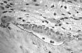
-
 Light micrograph of an osteoclast displaying typical distinguishing characteristics: a large cell with multiple nuclei and a "foamy" cytosol.
 -
 Light micrograph of osteoblasts, several displaying a prominent Golgi apparatus, actively synthesizing osteoid containing two osteocytes.
 -
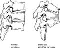 Collapse of vertebra on the right, normal on the left


Diagnosis

Osteoporosis often has no clear symptoms[112] in the early stages, making it difficult to detect without screening. However, you may experience signs such as unexplained back pain, loss of height, or a stooped posture as the condition progresses.[113] Osteoporosis can be diagnosed using conventional radiography and by measuring the bone mineral density (BMD).[114] The most popular method of measuring BMD is dual-energy X-ray absorptiometry.[115]
In addition to the detection of abnormal BMD, the diagnosis of osteoporosis requires investigations into potentially modifiable underlying causes; this may be done with blood tests. Depending on the likelihood of an underlying problem, investigations for cancer with metastasis to the bone, multiple myeloma, Cushing's disease, and other above-mentioned causes may be performed.[116]
Conventional radiography
Conventional radiography is useful, both by itself and in conjunction with CT or MRI, for detecting complications of osteopenia (reduced bone mass; pre-osteoporosis), such as fractures; for differential diagnosis of osteopenia; or follow-up examinations in specific clinical settings, such as soft tissue calcifications, secondary hyperparathyroidism, or osteomalacia in renal osteodystrophy. However, radiography is relatively insensitive to the detection of early disease and requires a substantial amount of bone loss (about 30%) to be apparent on X-ray images.[117][118]
The main radiographic features of generalized osteoporosis are cortical thinning and increased radiolucency. Frequent complications of osteoporosis are vertebral fractures, for which spinal radiography can help considerably in diagnosis and follow-up. Vertebral height measurements can be objectively made using plain-film X-rays by using several methods, such as height loss together with area reduction, particularly when looking at vertical deformity in T4-L4, or by determining a spinal fracture index that takes into account the number of vertebrae involved. Involvement of multiple vertebral bodies leads to kyphosis of the thoracic spine, leading to what is known as dowager's hump.[119][120]
Dual-energy X-ray
Dual-energy X-ray absorptiometry (DEXA scan) is considered the gold standard for the diagnosis of osteoporosis. Osteoporosis is diagnosed when the bone mineral density is less than or equal to 2.5 standard deviations below that of a young (30–40-year-old[7]:58), healthy adult women reference population. This is translated as a T-score. But because bone density decreases with age, more people become osteoporotic with increasing age.[7]:58 The World Health Organization has established the following diagnostic guidelines:[7][27]
| Category | T-score range | % young women |
|---|---|---|
| Normal | T-score ≥ −1.0 | 85% |
| Osteopenia | −2.5 < T-score < −1.0 | 14% |
| Osteoporosis | T-score ≤ −2.5 | 0.6% |
| Severe osteoporosis | T-score ≤ −2.5 with fragility fracture[27] |
The International Society for Clinical Densitometry takes the position that a diagnosis of osteoporosis in men under 50 years of age should not be made based on densitometric criteria alone. It also states that, for premenopausal women, Z-scores (comparison with age group rather than peak bone mass) rather than T-scores should be used, and the diagnosis of osteoporosis in such women also should not be made on the basis of densitometric criteria alone.[121]
Biomarkers
Chemical biomarkers are a useful tool in detecting bone degradation. The enzyme cathepsin K breaks down type-I collagen, an important constituent in bones. Prepared antibodies can recognize the resulting fragment, called a neoepitope, as a way to diagnose osteoporosis.[122] Increased urinary excretion of C-telopeptides, a type-I collagen breakdown product, also serves as a biomarker for osteoporosis.[123]
Other measuring tools
Quantitative computed tomography (QCT) differs from DXA in that it gives separate estimates of BMD for trabecular and cortical bone and reports precise volumetric mineral density in mg/cm3 rather than BMD's relative Z-score. Among QCT's advantages: it can be performed at axial and peripheral sites, can be calculated from existing CT scans without a separate radiation dose, is sensitive to change over time, can analyze a region of any size or shape, excludes irrelevant tissue such as fat, muscle, and air, and does not require knowledge of the patient's subpopulation in order to create a clinical score (e.g. the Z-score of all females of a certain age). Among QCT's disadvantages: it requires a high radiation dose compared to DXA, CT scanners are large and expensive, and because its practice has been less standardized than BMD, its results are more operator-dependent. Peripheral QCT has been introduced to improve upon the limitations of DXA and QCT.[114]
Quantitative ultrasound has many advantages in assessing osteoporosis. The modality is small, no ionizing radiation is involved, measurements can be made quickly and easily, and the cost of the device is low compared with DXA and QCT devices. The calcaneus is the most common skeletal site for quantitative ultrasound assessment because it has a high percentage of trabecular bone that is replaced more often than cortical bone, providing early evidence of metabolic change. Also, the calcaneus is fairly flat and parallel, reducing repositioning errors. The method can be applied to children, neonates, and preterm infants, just as well as to adults.[114] Some ultrasound devices can be used on the tibia.[124]
Screening
The U.S. Preventive Services Task Force (USPSTF) recommends that all women 65 years of age or older be screened by bone densitometry.[125] Additionally, they recommend screening younger women with risk factors.[125] There is insufficient evidence to make recommendations about the intervals for repeated screening and the appropriate age to stop screening.[126]
In men, the harm versus benefit of screening for osteoporosis is unknown.[125] Prescrire states that the need to test for osteoporosis in those who have not had a previous bone fracture is unclear.[127] The International Society for Clinical Densitometry suggest BMD testing for men 70 or older, or those who are indicated for risk equal to that of a 70‑year‑old.[128] A number of tools exist to help determine who is reasonable to test.[129]
Prevention
Lifestyle prevention of osteoporosis is, in many aspects, the inverse of the potentially modifiable risk factors.[130] As tobacco smoking and high alcohol intake have been linked with osteoporosis, smoking cessation and moderation of alcohol intake are commonly recommended as ways to help prevent it.[131]
In people with coeliac disease adherence to a gluten-free diet decreases the risk of developing osteoporosis[132] and increases bone density.[66] The diet must ensure optimal calcium intake (of at least one gram daily), and measuring vitamin D levels is recommended, and taking specific supplements if necessary.[132]
Osteoporosis can affect nearly 1 in 3 women, and the bone loss is the most rapid within the first 2–3 years after menopause. This can be prevented by menopause hormone therapy, or MHT, which is meant to prevent bone loss and the degradation of the bone microarchitecture and is noted to reduce the risk of fractures in bones by 20-30%. However, MHT has been linked to safety concerns, so it is not generally recommended.[133] As far as management goes with this potentially limiting disease, some practices can and should be implemented within the daily lifestyle. For example, it would be beneficial if the individual with osteoporosis refrained from consuming excess alcohol and avoided smoking.[134] These individuals should also be intentional about intaking an adequate amount of protein, calcium, and vitamin D. If the woman has an even higher risk of fracture, managing this may require therapy. Generally, the recommended treatment to prevent a decrease in bone mineral density is physical activity. Exercise is sometimes the best medicine. Resistance training is the most recommended method of physical activity, but that can come in multiple forms. High intensity and high impact training is shown to be extremely beneficial in improving bone health and the most effective in improving, maintaining bone density in the lower spine and femur. Although these types of exercises are safe for postmenopausal women, there may still be a need for supervision and precautionary measures.[135]
Nutrition
Studies of the benefits of supplementation with calcium and vitamin D are conflicting, possibly because most studies did not have people with low dietary intakes.[136] A 2018 review by the USPSTF found low-quality evidence that the routine use of calcium and vitamin D supplements (or both supplements together) did not reduce the risk of having an osteoporotic fracture in male and female adults living in the community who had no known history of vitamin D deficiency, osteoporosis, or a fracture.[137] The USPSTF does not recommend low dose supplementation (less than 1 g of calcium and 400 IU of vitamin D) in postmenopausal women as there does not appear to be a difference in fracture risk.[138] A 2015 review found little data that supplementation of calcium decreases the risk of fractures.[139] While some meta-analyses have found a benefit of vitamin D supplements combined with calcium for prevention of fractures, they did not find a benefit of vitamin D supplements (800 IU/day or less) alone.[140][141] Regarding adverse effects, supplementation does not appear to affect overall risk of death,[137][141] although calcium supplementation could potentially be associated with some increased risk of myocardial infarctions, stroke, kidney stones,[137] and gastrointestinal symptoms.[141] There is no evidence that supplementation before menopause can enhance bone mineral density.[142]
Vitamin K deficiency is also a risk factor for osteoporotic fractures.[143] The gene gamma-glutamyl carboxylase (GGCX) is dependent on vitamin K. Functional polymorphisms in the gene could attribute to variation in bone metabolism and BMD.[144] Vitamin K2 is also used as a means of treatment for osteoporosis and the polymorphisms of GGCX could explain the individual variation in the response to treatment of vitamin K.[145]
Dietary sources of calcium include dairy products, leafy greens, legumes, and beans.[146] There has been conflicting evidence about whether or not dairy is an adequate source of calcium to prevent fractures. The National Academy of Sciences recommends 1,000 mg of calcium for those aged 19–50, and 1,200 mg for those aged 50 and above.[147] A review of the evidence shows no adverse effect of higher protein intake on bone health.[50]
Physical exercise
Evidence suggests that exercise can help promote bone health in older people.[148] In particular, physical exercise can be beneficial for bone density in postmenopausal women,[149] and lead to a slightly reduced risk of a bone fracture (absolute difference 4%).[150] Weight bearing exercise has been found to cause an adaptive response in the skeleton,[151] promoting osteoblast activity and protecting bone density.[152] A position statement concluded that increased bone activity and weight-bearing exercises at a young age prevent bone fragility in adults.[153] Limitations in the available evidence hinder the production of detailed evidence-based exercise recommendations.[149] Some expert consensus guidance does exist.[154][155] International guidelines recommend multicomponent exercise tailored to individual needs that includes "balance and mobility training, paired with weight bearing exercise, progressive resistance training, and posture exercises" (generally accompanied by optimal nutrition).[156] Cycling and swimming are not considered weight-bearing exercise, and neither helps slow age-related bone loss (professional bicycle racing has a negative effect on bone density).[157] Risk of adverse events from the types of exercise usually considered appropriate for people with osteoporosis is generally low (though repeated forceful forward spinal bends are discouraged).[158]
For people who have had vertebral fractures, there is moderate-quality evidence that exercise is likely to improve physical performance, as well as some low-quality evidence suggesting that exercise may reduce pain and improve quality of life.[159]
Physical exercise prescription
Osteoporosis is a very prevalent disease in the elderly population, but not much is known about the optimal prescription and dosage of physical exercise to help prevent bone mineral loss. A lot of the focus around osteoporosis is prevention-oriented, rather than maintenance-focused, which should be the frontrunner when considering what approach to take. When prescribing exercise, an aspect to take into consideration is the individual's needs. This can be attained by conducting a pre-exercise evaluation or screening. Exercise should also be tailored to the individual and what works for them. Important things often overlooked when treating osteoporosis are muscle strength and maintenance of BMD, which should be incorporated into the program to optimize the benefits of exercise. This entails including exercises that focus on and improve muscle strength and exercises that focus on and improve skeletal strength or BMD, as these go hand in hand for reducing fall and fracture risk.[160] It's also important to reference the ACSM general training principle to better design a program for the individual. Which mode of exercise and dosage has been a recurring question for treating osteoporosis. Many studies have found that multimodal exercise programs improve factors related to osteoporosis. Factors include lower limb strength, balance, flexibility, and risk of falls.[161] Other modes of exercise have also proven to improve individuals with osteoporosis, some of these modes include weight-bearing, resistance specifically progressive resistance, and aerobic exercise. The recommendations for these types of exercises are as follows: weight-bearing exercise should be done 4–7 days a week, moderate to high intensity, activities should be multidirectional, and load should be more than the typical everyday load on bones.[160] Some examples of exercises are jumping, skipping, hopping, depth jumps, etc. Recommended dosage for progressive resistance training is 2 or more days a week, intensity (load) should start low and increase gradually.[160] Resistance training should focus on major muscle groups used for functional movements as well as muscles that have direct stress on bones susceptible to fracture. Considerations for resistance training are to teach proper lifting techniques and be careful with lifting weights above the head.[160] Lastly, aerobic exercise has minimal effect on preventing BMD loss unless done at a higher intensity or with a load like a weighted vest. Considerations with this mode are that this may cause a higher risk of falls or fractures.[160] Improvements can also be observed in other ways, such as decreased Timed-Up-and-Go, increased Sit-To-Stand, and increased One-Leg-Stance-Test. A study with a 12-week exercise intervention on postmenopausal osteoporotic women observed a 2.27 decrease in TUG times in their experimental group.[162] The overall thing to note when prescribing exercise for individuals with osteoporosis is to evaluate the individual's needs and then individualize their program with multiple exercise modalities that work for them, emphasizing increasing muscle strength as well as maintaining bone mass.
Physical therapy
People with osteoporosis are at higher risk of falls due to poor postural control, muscle weakness, and overall deconditioning.[163] Postural control is important to maintaining functional movements such as walking and standing. Physical therapy may be an effective way to address postural weakness that may result from vertebral fractures, which are common in people with osteoporosis. Physical therapy treatment plans for people with vertebral fractures include balance training, postural correction, trunk and lower extremity muscle strengthening exercises, and moderate-intensity aerobic physical activity.[159] The goal of these interventions are to regain normal spine curvatures, increase spine stability, and improve functional performance.[159] Physical therapy interventions were also designed to slow the rate of bone loss through home exercise programs.[163]
Whole body vibration therapy has also been suggested as a physical therapy intervention. Moderate to low-quality evidence indicates that whole body vibration therapy may reduce the risk of falls.[164] There are conflicting reviews as to whether vibration therapy improves bone mineral density.[164][165]
People with osteoporosis are at higher risk of falls due to poor posture, muscle weakness, and deconditioning, making physical therapy important for improving balance and function. Treatment includes balance training, postural correction, strengthening, and aerobic exercise to improve stability and slow bone mineral density (BMD) loss. Regular weight-bearing and resistance exercise also promotes bone formation, making it an effective non-drug approach for prevention and management.[166]
Physical therapy can aid in the overall prevention in the development of osteoporosis through therapeutic exercise. Prescribed amounts of mechanical loading or increased forces on the bones promote bone formation and vascularization in various ways, therefore offering a preventative measure that is not reliant on drugs. Specific exercise interacts with the body's hormones and signaling pathways, which encourages the maintenance of a healthy skeleton.[167]
Hormone therapy
Reduced estrogen levels increase the risk of osteoporosis, so hormone replacement therapy when women reach the menopause may reduce the incidence of osteoporosis. A more natural way of restoring hormone levels in postmenopausal women includes participating in specific forms of exercise. Weight-bearing exercises and resistance training exercises such as squats with weights, step-ups, lunges, stair climbing, and even jogging can elicit hormone responses that are advantageous for post-menopausal women living with osteoporosis. These exercises result in the release of growth hormone and Insulin-like growth factor-1 or IGF-1 which participate in bone remodeling.[168] Stress is applied to the bones, thus activating osteoblast, which are cells that form new bones and grow and heal existing bones[169] while restoring hormones that increase bone density. Resistance training exercises, like weightlifting, can lead to a brief increase in anabolic hormones, like testosterone, which aid in muscle and bone strength.[170]
The increase in mechanical tension during resistance exercise will likely help stimulate the production of Insulin-like growth factors in the bone, but at a greater extent. Post-menopausal women experience a reduction of estrogen, which is essential for bone density, so these exercise-induced hormonal enhancements can counteract the loss of bone mineral density in the most critical areas, like the lumbar spine and the femoral neck.[171] Research suggests that regular resistance training accompanied with weight-bearing activities help reduce the progression of osteoporosis and risk of fracture.
Management
Lifestyle Changes
Weight-bearing endurance exercise and/or exercises to strengthen muscles improve bone strength in those with osteoporosis.[150][172] Aerobics, weight bearing, and resistance exercises all maintain or increase BMD in postmenopausal women.[150][173] Daily intake of calcium and vitamin D is recommended for postmenopausal women.[173] Fall prevention can help prevent osteoporosis complications. There is some evidence for hip protectors specifically among those who are in care homes.[174]
Pharmacologic therapy
The US National Osteoporosis Foundation recommends pharmacologic treatment for patients with hip or spine fracture thought to be related to osteoporosis, those with BMD 2.5 SD or more below the young normal mean (T-score -2.5 or below), and those with BMD between 1 and 2.5 SD below normal mean whose 10-year risk, using FRAX, for hip fracture is equal or more than 3%.[175] Bisphosphonates are useful in decreasing the risk of future fractures in those who have already sustained a fracture due to osteoporosis.[8][9][131][176] This benefit is present when taken for three to four years.[177][178] They do not appear to change the overall risk of death.[11] Tentative evidence does not support the use of bisphosphonates as a standard treatment for secondary osteoporosis in children.[178] Different bisphosphonates have not been directly compared; therefore, it is unknown if one is better than another.[131] Fracture risk reduction is between 25 and 70%, depending on the bone involved.[131] There are concerns of atypical femoral fractures and osteonecrosis of the jaw with long-term use, but these risks are low.[131][179] With evidence of little benefit when used for more than three to five years and in light of the potential adverse events, it may be appropriate to stop treatment after this time.[177] One medical organization recommends that after five years of medications by mouth or three years of intravenous medication among those at low risk, bisphosphonate treatment can be stopped.[180][181] In those at higher risk, they recommend up to ten years of medication by mouth or six years of intravenous treatment.[180]
The goal of osteoporosis management is to prevent osteoporotic fractures, but for those who have sustained one already it is more urgent to prevent a secondary fracture.[182] That is because patients with a fracture are more likely to experience a recurrent fracture, with a marked increase in morbidity and mortality compared.[182] Among the five bisphosphonates, no significant differences were found for a secondary fracture for all fracture endpoints combined.[182] That being said, alendronate was identified as the most efficacious for secondary prevention of vertebral and hip fractures, while zoledronate showed better performance for nonvertebral non-hip fracture prevention.[182] There is concern that many people do not receive appropriate pharmacological therapy after a low-impact fracture.[183]
For those with osteoporosis but who have not had a fracture, evidence does not support a reduction in fracture risk with risedronate[9] or etidronate.[10] Alendronate decreases fractures of the spine but does not have any effect on other types of fractures.[8] Half stop their medications within a year.[184] When on treatment with bisphosphonates rechecking bone mineral density is not needed.[181] There is tentative evidence of benefit in males with osteoporosis.[185]
Fluoride supplementation does not appear to be effective in postmenopausal osteoporosis, as even though it increases bone density, it does not decrease the risk of fractures.[186][187]
Teriparatide (a recombinant parathyroid hormone) is effective in treatment of women with postmenopausal osteoporosis.[188][176] Some evidence also indicates strontium ranelate is effective in decreasing the risk of vertebral and nonvertebral fractures in postmenopausal women with osteoporosis.[189] Hormone replacement therapy, while effective for osteoporosis, is only recommended in women who also have menopausal symptoms.[131] It is not recommended for osteoporosis by itself.[181] Raloxifene, while effective in decreasing vertebral fractures, does not affect the risk of nonvertebral fracture.[131] And while it reduces the risk of breast cancer, it increases the risk of blood clots and strokes.[131] While denosumab is effective at preventing fractures in women,[131] there is not clear evidence of benefit in males.[185] In hypogonadal men, testosterone has been shown to improve bone quantity and quality, but, as of 2008, no studies evaluated its effect on fracture risk or in men with normal testosterone levels.[65] Calcitonin, while once recommended, is no longer recommended due to the associated risk of cancer and questionable effect on fracture risk.[190] Alendronic acid/colecalciferol can be taken to treat this condition in post-menopausal women.[191]
Romosozumab (sold under the brand name Evenity) is a monoclonal antibody against sclerostin. Romosozumab is usually reserved for patients with very high fracture risk and is the only available drug therapy for osteoporosis that leads to simultaneous inhibition of bone resorption together with an anabolic effect.[192][193]
Certain medications like alendronate, etidronate, risedronate, raloxifene, and strontium ranelate can help to prevent osteoporotic fragility fractures in postmenopausal women with osteoporosis.[194] Tentative evidence suggests that Chinese herbal medicines may have potential benefits on bone mineral density.[195]
Prognosis
| WHO category | Age 50–64 | Age > 64 | Overall |
|---|---|---|---|
| Normal | 5.3 | 9.4 | 6.6 |
| Osteopenia | 11.4 | 19.6 | 15.7 |
| Osteoporosis | 22.4 | 46.6 | 40.6 |
Although people with osteoporosis have increased mortality due to the complications of fracture, the fracture itself is rarely lethal.
Hip fractures can lead to decreased mobility and additional risks of numerous complications (such as deep venous thrombosis and/or pulmonary embolism, and pneumonia). The six-month mortality rate for those aged 50 and above following hip fracture was found to be around 13.5%, with a substantial proportion (almost 13%) needing total assistance to mobilize after a hip fracture.[197]
Vertebral fractures, while having a smaller impact on mortality, can lead to severe chronic pain of neurogenic origin, which can be hard to control, as well as deformity. Though rare, multiple vertebral fractures can lead to such severe hunchback (kyphosis), the resulting pressure on internal organs can impair one's ability to breathe.
Apart from the risk of death and other complications, osteoporotic fractures are associated with a reduced health-related quality of life.[198]
The condition is responsible for millions of fractures annually, mostly involving the lumbar vertebrae, hip, and wrist. Fragility fractures of ribs are also common in men.
Fractures
Hip fractures are responsible for the most serious consequences of osteoporosis. In the United States, more than 250,000 hip fractures annually are attributable to osteoporosis.[199] A 50-year-old white woman is estimated to have a 17.5% lifetime risk of fracture of the proximal femur. The incidence of hip fractures increases each decade from the sixth through the ninth for both women and men across all populations. The highest incidence is found among men and women aged 80 or older.[200]
Between 35 and 50% of all women over 50 had at least one vertebral fracture. In the United States, 700,000 vertebral fractures occur annually, but only about a third are recognized. In a series of 9704 women aged 68.8 on average studied for 15 years, 324 had already sustained a vertebral fracture at entry into the study, and 18.2% developed a vertebral fracture, but that risk rose to 41.4% in women who had a previous vertebral fracture.[201]
In the United States, 250,000 wrist fractures annually are attributable to osteoporosis.[199] Wrist fractures are the third most common type of osteoporotic fractures. The lifetime risk of sustaining a Colles' fracture is about 16% for white women. By the time women reach age 70, about 20% have had at least one wrist fracture.[200]
Fragility fractures of the ribs are common in men as young as age 35. These are often overlooked as signs of osteoporosis, as these men are often physically active and develop the fracture in the course of physical activity, such as falling while water skiing or jet skiing.
Epidemiology
| This medicine needs to be updated. Please update this medicine to reflect recent events or newly available information. (December 2020) |

Osteoporosis becomes more common with age, especially after 50 years (its prevalence rises from about 2% at 50 years to almost 50% by the age of 80).[203] It affects women more than men due to the sharp fall in estrogen production that follows menopause.[203] Globally, it is estimated that 21.2% of women and 6.3% of men over the age of 50 have osteoporosis, corresponding to a total of around 500 million people worldwide.[204] About 15% of Caucasians in their 50s and 70% of those over 80 are affected.[12] In the developed world, depending on the method of diagnosis, 2% to 8% of males and 9% to 38% of females are affected.[13] Rates of disease in the developing world are unclear.[14]
From the age of 50 onwards, fractures (including hip fractures) are roughly twice as common in women than in men.[205] A 60-year-old woman has a 44% chance of experiencing a fracture in her lifetime, whereas the lifetime risk for a 60-year-old man is only 25%.[206] Such differences can be attributed to the increased risk of osteoporosis due to decreased estrogen levels following menopause.[206]
In 2019,[207] up to 37 million fragility fractures linked to osteoporosis were thought to occur in people over the age of 55 worldwide.[204] Globally, 1 in 3 women and 1 in 5 men over the age of 50 will have an osteoporotic fracture.[204] Data from the United States shows a decrease in osteoporosis within the general population and in white women, from 18% in 1994 to 10% in 2006.[208] White and Asian people are at greater risk.[2] People of African descent are at a decreased risk of fractures due to osteoporosis, although they have the highest risk of death following an osteoporotic fracture.[208]
It has been shown that latitude affects the risk of osteoporotic fracture.[202] Areas of higher latitude such as Northern Europe receive less Vitamin D through sunlight compared to regions closer to the equator, and consequently have higher fracture rates in comparison to lower latitudes.[202] For example, Swedish men and women have a 13% and 28.5% risk of hip fracture by age 50, respectively, whereas this risk is only 1.9% and 2.4% in Chinese men and women.[208] Diet may also be a factor that is responsible for this difference, as vitamin D, calcium, magnesium, and folate are all linked to bone mineral density.[209]
There is also an association between Celiac Disease and increased risk of osteoporosis.[210] In studies with premenopausal females and males, there was a correlation between Celiac Disease and osteoporosis and osteopenia.[210] Celiac Disease can decrease absorption of nutrients in the small intestine such as calcium, and a gluten-free diet can help people with Celiac Disease to revert to normal absorption in the gut.[211]
About 22 million women and 5.5 million men in the European Union had osteoporosis in 2010.[15] In the United States in 2010, about 8 million women and one to 2 million men had osteoporosis.[13][16] This places a large economic burden on the healthcare system due to costs of treatment, long-term disability, and loss of productivity in the working population. The EU spends 37 billion euros per year on healthcare costs related to osteoporosis, and the US spends an estimated US$19 billion annually on related healthcare costs.[212]
History
Research on age-related reductions in bone density goes back to the early 1800s. French pathologist Jean Lobstein coined the term osteoporosis.[17] The American endocrinologist Fuller Albright linked osteoporosis with the postmenopausal state.[213]
Anthropologists have studied skeletal remains that showed loss of bone density and associated structural changes that were linked to a chronic malnutrition in the agricultural area in which these individuals lived. "It follows that the skeletal deformation may be attributed to their heavy labor in agriculture as well as to their chronic malnutrition", causing the osteoporosis seen when radiographs of the remains were made.[214]
See also
- Fracture Risk Assessment Tool (FRAX)
- Spaceflight osteopenia
- World Osteoporosis Day
References
- ↑ https://my.clevelandclinic.org/health/body/24871-osteoblasts-and-osteoclasts
- ↑ 2.0 2.1 2.2 2.3 2.4 2.5 2.6 "Handout on Health: Osteoporosis". August 2014. https://www.niams.nih.gov/health-topics/osteoporosis.
- ↑ "Osteoporosis: screening, prevention, and management". The Medical Clinics of North America 99 (3): 587–606. May 2015. doi:10.1016/j.mcna.2015.01.010. PMID 25841602. https://zenodo.org/record/1259215.
- ↑ NIAMS Science Communications and Outreach Branch (2017-04-07). "Osteoporosis" (in en). https://www.niams.nih.gov/health-topics/osteoporosis.
- ↑ "Menopause and Bone Loss" (in en). 2022-01-24. https://www.endocrine.org/patient-engagement/endocrine-library/menopause-and-bone-loss.
- ↑ "Clinical Challenges: Managing Osteoporosis in Male Hypogonadism" (in en). Ziff Davis. 2018-06-04. https://www.medpagetoday.com/clinical-challenges/aace-osteoporosis/73256.
- ↑ 7.00 7.01 7.02 7.03 7.04 7.05 7.06 7.07 7.08 7.09 7.10 7.11 7.12 7.13 7.14 7.15 Prevention and management of osteoporosis. World Health Organization Technical Report Series. 921. World Health Organization. 2003. pp. 1–164, back cover. ISBN 978-92-4-120921-2.
- ↑ 8.0 8.1 8.2 "Alendronate for the primary and secondary prevention of osteoporotic fractures in postmenopausal women". The Cochrane Database of Systematic Reviews (1). January 2008. doi:10.1002/14651858.CD001155.pub2. PMID 18253985.
- ↑ 9.0 9.1 9.2 "Risedronate for the primary and secondary prevention of osteoporotic fractures in postmenopausal women". The Cochrane Database of Systematic Reviews 2022 (7). May 2022. doi:10.1002/14651858.CD004523.pub4. PMID 35502787.
- ↑ 10.0 10.1 Wells, George A.; Hsieh, Shu-Ching; Peterson, Joan; Zheng, Carine; Kelly, Shannon E.; Shea, Beverley; Tugwell, Peter (2024-04-09). "Etidronate for the primary and secondary prevention of osteoporotic fractures in postmenopausal women". The Cochrane Database of Systematic Reviews 2024 (4). doi:10.1002/14651858.CD003376.pub4. ISSN 1469-493X. PMID 38591743.
- ↑ 11.0 11.1 "Association Between Drug Treatments for Patients With Osteoporosis and Overall Mortality Rates: A Meta-analysis". JAMA Internal Medicine 179 (11): 1491–1500. August 2019. doi:10.1001/jamainternmed.2019.2779. PMID 31424486.
- ↑ 12.0 12.1 "Chronic rheumatic conditions". https://www.who.int/chp/topics/rheumatic/en/.
- ↑ 13.0 13.1 13.2 13.3 "Estimating prevalence of osteoporosis: examples from industrialized countries". Archives of Osteoporosis 9 (1). 2014. doi:10.1007/s11657-014-0182-3. PMID 24847682.
- ↑ 14.0 14.1 "Osteoporosis in developing countries". Best Practice & Research. Clinical Rheumatology 22 (4): 693–708. August 2008. doi:10.1016/j.berh.2008.04.002. PMID 18783745.
- ↑ 15.0 15.1 "Osteoporosis in the European Union: a compendium of country-specific reports". Archives of Osteoporosis 8 (1–2). 2013. doi:10.1007/s11657-013-0137-0. PMID 24113838.
- ↑ 16.0 16.1 "The clinical epidemiology of male osteoporosis: a review of the recent literature". Clinical Epidemiology 7: 65–76. 2015. doi:10.2147/CLEP.S40966. PMID 25657593.
- ↑ 17.0 17.1 Grob, Gerald N. (2014). Aging Bones: A Short History of Osteoporosis. JHU Press. p. 5. ISBN 978-1-4214-1318-1.
- ↑ 18.0 18.1 "Osteoporosis" (in en). National Institute of Arthritis and Musculoskeletal and Skin Diseases, US National Institutes of Health. 1 December 2022. https://www.niams.nih.gov/health-topics/osteoporosis.
- ↑ Harrison's principles of internal medicine. (Twentieth ed.). New York: McGraw-Hill Education. 2018-02-06. ISBN 978-1-259-64404-7. OCLC 990065894.
- ↑ "Vertebral compression fractures in the elderly". American Family Physician 69 (1): 111–116. January 2004. PMID 14727827. http://www.aafp.org/afp/2004/0101/p111.html. Retrieved 31 March 2011.
- ↑ "Identification of prevalent vertebral fractures using Vertebral Fracture Assessment (VFA) in asymptomatic postmenopausal women: A systematic review and meta-analysis". Bone 136. July 2020. doi:10.1016/j.bone.2020.115358. PMID 32268210.
- ↑ "Osteoporotic compression fractures of the spine; current options and considerations for treatment". The Spine Journal 6 (5): 479–487. 2006. doi:10.1016/j.spinee.2006.04.013. PMID 16934715.
- ↑ "Changing trends in the mortality rate at 1-year post hip fracture - a systematic review". World Journal of Orthopedics 10 (3): 166–175. March 2019. doi:10.5312/wjo.v10.i3.166. PMID 30918799.
- ↑ Susan Ott (October 2009). "Fracture Risk Calculator". http://courses.washington.edu/bonephys/FxRiskCalculator.html.
- ↑ "A New Fracture Risk Assessment Tool (FREM) Based on Public Health Registries". Journal of Bone and Mineral Research 33 (11): 1967–1979. November 2018. doi:10.1002/jbmr.3528. PMID 29924428.
- ↑ "Administrative healthcare data applied to fracture risk assessment". Osteoporosis International 30 (3): 565–571. March 2019. doi:10.1007/s00198-018-4780-6. PMID 30554259.
- ↑ 27.0 27.1 27.2 Assessment of fracture risk and its application to screening for postmenopausal osteoporosis. World Health Organization Technical Report Series. 843. World Health Organization. 1994. pp. 1–129. ISBN 978-92-4-120843-7.
- ↑ "Will my patient fall?". JAMA 297 (1): 77–86. 2007. doi:10.1001/jama.297.1.77. PMID 17200478.
- ↑ "Osteoporosis - Symptoms and causes" (in en). https://www.mayoclinic.org/diseases-conditions/osteoporosis/symptoms-causes/syc-20351968.
- ↑ "An overview and management of osteoporosis". European Journal of Rheumatology 4 (1): 46–56. March 2017. doi:10.5152/eurjrheum.2016.048. PMID 28293453.
- ↑ "Risk factors for low bone mass in healthy 40–60 year old women: a systematic review of the literature". Osteoporosis International 20 (1): 1–21. January 2009. doi:10.1007/s00198-008-0643-x. PMID 18523710.
- ↑ "Racial and Ethnic Disparities in Bone Health and Outcomes in the United States". Journal of Bone and Mineral Research 36 (10): 1881–1905. 2021. doi:10.1002/jbmr.4417. PMID 34338355.
- ↑ "6.6 Exercise, Nutrition, Hormones, and Bone Tissue". Anatomy & Physiology. Openstax CNX. 2013. ISBN 978-1-938168-13-0. http://cnx.org/contents/FPtK1zmh@6.27:g-vsB2Y2@4/Exercise-Nutrition-Hormones-an.
- ↑ "Novel insights in the regulation and mechanism of androgen action on bone". Current Opinion in Endocrinology, Diabetes and Obesity 20 (3): 240–44. 2013. doi:10.1097/MED.0b013e32835f7d04. PMID 23449008.
- ↑ "Testosterone and the male skeleton: a dual mode of action". Journal of Osteoporosis 2011: 1–7. 2011. doi:10.4061/2011/240328. PMID 21941679.
- ↑ Melton LJ (2003). "Epidemiology worldwide". Endocrinol. Metab. Clin. North Am. 32 (1): v, 1–13. doi:10.1016/S0889-8529(02)00061-0. PMID 12699289.
- ↑ Yang, Tie-Lin; Shen, Hui; Liu, Anqi; Dong, Shan-Shan; Zhang, Lei; Deng, Fei-Yan; Zhao, Qi; Deng, Hong-Wen (2020). "A road map for understanding molecular and genetic determinants of osteoporosis" (in en). Nature Reviews Endocrinology 16 (2): 91–103. doi:10.1038/s41574-019-0282-7. ISSN 1759-5037. https://www.nature.com/articles/s41574-019-0282-7.
- ↑ "History of fractures as predictor of subsequent hip and nonhip fractures among older Mexican Americans". Journal of the National Medical Association 99 (4): 412–418. 2007. PMID 17444431.
- ↑ 39.0 39.1 Brian K Alldredge; Koda-Kimble, Mary Anne; Young, Lloyd Y.; Wayne A Kradjan; B. Joseph Guglielmo (2009). Applied therapeutics: the clinical use of drugs. Philadelphia: Wolters Kluwer Health/Lippincott Williams & Wilkins. pp. 101–103. ISBN 978-0-7817-6555-8.
- ↑ 40.0 40.1 "Osteoporosis and its management". BMJ 333 (7581): 1251–1256. December 2006. doi:10.1136/bmj.39050.597350.47. PMID 17170416.
- ↑ "Association between alcohol consumption and both osteoporotic fracture and bone density". Am J Med 121 (5): 406–418. 2008. doi:10.1016/j.amjmed.2007.12.012. PMID 18456037.
- ↑ Nieves JW (2005). "Osteoporosis: the role of micronutrients". Am J Clin Nutr 81 (5): 1232S–1239S. doi:10.1093/ajcn/81.5.1232. PMID 15883457.
- ↑ "Calcium and vitamin d supplementation in men". Journal of Osteoporosis 2011: 1–6. 2011. doi:10.4061/2011/875249. PMID 21876835.
- ↑ "Association between electronic cigarette use and fragility fractures among US adults" (in en). American Journal of Medicine Open 1-6. January 2021. doi:10.1016/j.ajmo.2021.100002. ISSN 2667-0364. PMID 39036626.
- ↑ "Cigarette smoking and bone mineral density in older men and women". American Journal of Public Health 83 (9): 1265–1270. September 1993. doi:10.2105/AJPH.83.9.1265. PMID 8363002.
- ↑ "Smoking and fracture risk: a meta-analysis". Osteoporosis International 16 (2): 155–162. February 2005. doi:10.1007/s00198-004-1640-3. PMID 15175845.
- ↑ Wong, Peter K. K.; Christie, Jemma J.; Wark, John D. (September 2007). "The effects of smoking on bone health". Clinical Science 113 (5): 233–241. doi:10.1042/CS20060173. PMID 17663660.
- ↑ Ilich, Jasminka Z.; Kerstetter, Jane E. (December 2000). "Nutrition in Bone Health Revisited: A Story Beyond Calcium". Journal of the American College of Nutrition 19 (6): 715–737. doi:10.1080/07315724.2000.10718070. PMID 11194525.
- ↑ "Ratio of n−6 to n−3 fatty acids and bone mineral density in older adults: the Rancho Bernardo Study". Am J Clin Nutr 81 (4): 934–38. 2005. doi:10.1093/ajcn/81.4.934. PMID 15817874.
- ↑ 50.0 50.1 "Dietary protein and bone health: a systematic review and meta-analysis from the National Osteoporosis Foundation". The American Journal of Clinical Nutrition 105 (6): 1528–1543. June 2017. doi:10.3945/ajcn.116.145110. PMID 28404575.
- ↑ "Protein intake and bone health: an umbrella review of systematic reviews for the evidence-based guideline of the German Nutrition Society". Osteoporosis International 34 (8): 1335–1353. August 2023. doi:10.1007/s00198-023-06709-7. PMID 37126148.
- ↑ "Influence of muscle strength on bone strength during childhood and adolescence". Hormone Research 45 (Suppl. 1): 63–66. 1996. doi:10.1159/000184834. PMID 8805035.
- ↑ "Bone, body weight, and weight reduction: what are the concerns?". J. Nutr. 136 (6): 1453–1456. 1 June 2006. doi:10.1093/jn/136.6.1453. PMID 16702302.
- ↑ Pollock, Noel; Grogan, Claire; Perry, Mark; Pedlar, Charles; Cooke, Karl; Morrissey, Dylan; Dimitriou, Lygeri (October 2010). "Bone-Mineral Density and Other Features of the Female Athlete Triad in Elite Endurance Runners: A Longitudinal and Cross-Sectional Observational Study". International Journal of Sport Nutrition and Exercise Metabolism 20 (5): 418–426. doi:10.1123/ijsnem.20.5.418. PMID 20975110. http://journals.humankinetics.com/ijsnem.
- ↑ "Nutritional and exercise-related determinants of bone density in elite female runners". Osteoporosis International 15 (8): 611–618. 2004. doi:10.1007/s00198-004-1589-2. PMID 15048548.
- ↑ "Low bone mass and high bone turnover in male long distance runners". The Journal of Clinical Endocrinology and Metabolism 77 (3): 770–775. 1993. doi:10.1210/jcem.77.3.8370698. PMID 8370698.
- ↑ "Bone metabolism in endurance trained athletes: A comparison to population-based controls based on DXA, SXA, quantitative ultrasound, and biochemical markers". Calcified Tissue International 61 (6): 448–454. 1997. doi:10.1007/s002239900366. PMID 9383270.
- ↑ "Bone mineral density and serum testosterone in chronically trained, high mileage 40–55 year old male runners". British Journal of Sports Medicine 34 (4): 273–278. 2000. doi:10.1136/bjsm.34.4.273. PMID 10953900.
- ↑ "Environmental exposure to cadmium, forearm bone density, and risk of fractures: prospective population study. Public Health and Environmental Exposure to Cadmium (PheeCad) Study Group". Lancet 353 (9159): 1140–1144. 1999. doi:10.1016/S0140-6736(98)09356-8. PMID 10209978.
- ↑ "Colas, but not other carbonated beverages, are associated with low bone mineral density in older women: The Framingham Osteoporosis Study". Am. J. Clin. Nutr. 84 (4): 936–942. 2006. doi:10.1093/ajcn/84.4.936. PMID 17023723.
- ↑ American Academy of Pediatrics Committee on School Health (2004). "Soft drinks in schools". Pediatrics 113 (1 Pt 1): 152–54. doi:10.1542/peds.113.1.152. PMID 14702469.
- ↑ "Proton-pump inhibitors and risk of fractures: an update meta-analysis". Osteoporosis International 27 (1): 339–347. January 2016. doi:10.1007/s00198-015-3365-x. PMID 26462494.
- ↑ 63.0 63.1 63.2 63.3 63.4 Simonelli, C (July 2006). "ICSI Health Care Guideline: Diagnosis and Treatment of Osteoporosis, 5th edition" (PDF). Institute for Clinical Systems Improvement. http://www.icsi.org/osteoporosis/diagnosis_and_treatment_of_osteoporosis__3.html.
- ↑ 64.00 64.01 64.02 64.03 64.04 64.05 64.06 64.07 64.08 64.09 64.10 64.11 Osteoporosis at eMedicine
- ↑ 65.0 65.1 65.2 Ebeling PR (2008). "Clinical practice. Osteoporosis in men". N Engl J Med 358 (14): 1474–1482. doi:10.1056/NEJMcp0707217. PMID 18385499.
- ↑ 66.0 66.1 "Management of endocrine disease: Secondary osteoporosis: pathophysiology and management". Eur J Endocrinol 173 (3): R131–151. Sep 2015. doi:10.1530/EJE-15-0118. PMID 25971649.
- ↑ 67.0 67.1 67.2 Henwood, Maria J.; Binkovitz, Larry (January 2009). "Update on pediatric bone health". The Journal of the American Osteopathic Association 109 (1): 5–12. PMID 19193819.
- ↑ "The role of calcium in human aging". Clin Nutr Res 4 (1): 1–8. Jan 2015. doi:10.7762/cnr.2015.4.1.1. PMID 25713787.
- ↑ "Chronic Kidney Disease". https://www.lecturio.com/concepts/chronic-kidney-disease/.
- ↑ "Often seen, rarely recognized: mast cell activation disease--a guide to diagnosis and therapeutic options". The American Journal of the Medical Science 48 (3): 190–201. 2016. doi:10.3109/07853890.2016.1161231. PMID 27012973.
- ↑ "Mast cell activation syndrome: An up-to-date review of literature". World J Clin Pediatr 13 (2). 2024-06-09. doi:10.5409/wjcp.v13.i2.92813. PMID 38948000.
- ↑ "Mast cell activation disease: a concise practical guide for diagnostic workup and therapeutic options". Dtsch Med Wochenschr 129 (30): 1523–34. Jul 2014. doi:10.1055/s-0034-1370055. PMID 24801454.
- ↑ "Three cases of multicentric carpotarsal osteolysis syndrome: a case series". BMC Medical Genetics 19 (1). September 2018. doi:10.1186/s12881-018-0682-x. PMID 30208859.
- ↑ "Multicentric Osteolysis, Nodulosis, and Arthropathy in two unrelated children with matrix metalloproteinase 2 variants: Genetic-skeletal correlations". Bone Reports 15. December 2021. doi:10.1016/j.bonr.2021.101106. PMID 34307793.
- ↑ Nunes, Mark E. (1993). "Hypophosphatasia". GeneReviews®. University of Washington, Seattle. https://www.ncbi.nlm.nih.gov/books/NBK1150/.
- ↑ "Hypophosphatasia Case Studies: Dangers of Misdiagnosis". http://www.hypophosphatasia.com/hcp/dangers-of-misdiagnosis.
- ↑ "Osteoporosis in Parkinson's disease". Parkinsonism & Related Disorders 15 (5): 339–346. 2009. doi:10.1016/j.parkreldis.2009.02.009. PMID 19346153.
- ↑ "Mitochondria, calcium and cell death: A deadly triad in neurodegeneration". Biochimica et Biophysica Acta (BBA) - Bioenergetics 1787 (5): 335–344. 2009. doi:10.1016/j.bbabio.2009.02.021. PMID 19268425.
- ↑ Bone and Tooth Society of Great Britain; National Osteoporosis Society; Royal College of Physicians (2003). Glucocorticoid-induced Osteoporosis. London: Royal College of Physicians of London. ISBN 978-1-86016-173-5. http://bookshop.rcplondon.ac.uk/contents/pub89-a953a6c0-06c0-46d8-b79a-e951536d9070.pdf. Retrieved 3 October 2011.
- ↑ "Calcium and vitamin D for corticosteroid-induced osteoporosis". The Cochrane Database of Systematic Reviews 1998 (2). 1998-04-27. doi:10.1002/14651858.cd000952. PMID 10796394.
- ↑ "Prevention and treatment strategies for glucocorticoid-induced osteoporotic fractures". Clinical Rheumatology 26 (2): 144–153. February 2007. doi:10.1007/s10067-006-0315-1. PMID 16670825.
- ↑ "Anti-epileptic medication and bone health". Osteoporosis International 18 (2): 129–142. 2007. doi:10.1007/s00198-006-0185-z. PMID 17091219.
- ↑ "Heparin and osteoporosis during pregnancy: 2002 update". Lupus 11 (10): 680–682. 2002. doi:10.1191/0961203302lu262oa. PMID 12413068.
- ↑ "Long-term proton pump inhibitor therapy and risk of hip fracture". JAMA 296 (24): 2947–2953. 2006. doi:10.1001/jama.296.24.2947. PMID 17190895.
- ↑ "Effects of thiazolidinediones on bone loss and fracture". Annals of Pharmacotherapy 41 (12): 2014–2018. 2007. doi:10.1345/aph.1K286. PMID 17940125.
- ↑ Hardcastle, Sarah A. (May 2022). "Pregnancy and Lactation Associated Osteoporosis". Calcified Tissue International 110 (5): 531–545. doi:10.1007/s00223-021-00815-6. PMID 33620518.
- ↑ Lujano-Negrete, Abraham Yair; Rodríguez-Ruiz, Martha Cecilia; Skinner-Taylor, Cassandra Michele; Perez-Barbosa, Lorena; Cardenas de la Garza, Jesus Alberto; García-Hernández, Pedro Alberto; Espinosa-Banuelos, Luis Gerardo; Gutierrez-Leal, Luz Fernanda et al. (December 2022). "Bone metabolism and osteoporosis during pregnancy and lactation". Archives of Osteoporosis 17 (1): 36. doi:10.1007/s11657-022-01077-x. PMID 35182221.
- ↑ Phillips, A. J.; Ostlere, S. J.; Smith, R. (May 2000). "Pregnancy-Associated Osteoporosis: Does the Skeleton Recover?". Osteoporosis International 11 (5): 449–454. doi:10.1007/s001980070113. PMID 10912848.
- ↑ Komnos, George A.; Paridis, Dionyssios; Donoudis, Christos; Sotiriou, Sotirios (30 January 2024). "Gestational osteoporosis: Myth or true?". International Journal of Biology and Pharmacy Research Updates 3 (1): 001–011. doi:10.53430/ijbpru.2024.3.1.0040.
- ↑ Rizzoli, R; Bonjour, JP (August 1996). "Pregnancy-associated osteoporosis". The Lancet 348 (9024): 403. doi:10.1016/S0140-6736(05)65018-0.
- ↑ 91.0 91.1 Latimer B (2005). "The perils of being bipedal". Ann Biomed Eng 33 (1): 3–6. doi:10.1007/s10439-005-8957-8. PMID 15709701.
- ↑ 92.0 92.1 92.2 Cotter M (2011). "Human evolution and osteoporosis-related spinal fractures". PLOS ONE 6 (10). doi:10.1371/journal.pone.0026658. PMID 22028933. Bibcode: 2011PLoSO...626658C.
- ↑ "Calcium in evolutionary perspective". Am. J. Clin. Nutr. 54 (1 Suppl): 281S–287S. 1991. doi:10.1093/ajcn/54.1.281S. PMID 2053574.
- ↑ Bono, Christopher M.; Einhorn, Thomas A. (2005). "Overview of osteoporosis: Pathophysiology and determinants of bone strength". The Aging Spine. European Spine Journal. 12. pp. 8–14. doi:10.1007/3-540-27376-X_3. ISBN 978-3-540-24408-0.
- ↑ 95.0 95.1 95.2 "Bone three-dimensional microstructural features of the common osteoporotic fracture sites". World Journal of Orthopedics 5 (4): 486–495. September 2014. doi:10.5312/wjo.v5.i4.486. PMID 25232524.
- ↑ "Bone mechanical properties and changes with osteoporosis". Injury 47 (Suppl 2): S11–S20. June 2016. doi:10.1016/s0020-1383(16)47003-8. PMID 27338221.
- ↑ 97.0 97.1 97.2 "Whole bone mechanics and mechanical testing". Veterinary Journal 177 (1): 8–17. July 2008. doi:10.1016/j.tvjl.2007.09.012. PMID 17986396.
- ↑ 98.0 98.1 98.2 98.3 98.4 "The fracture mechanics of human bone: influence of disease and treatment". BoneKEy Reports 4: 743. 2015-09-02. doi:10.1038/bonekey.2015.112. PMID 26380080.
- ↑ "Bone quality changes associated with aging and disease: a review". Annals of the New York Academy of Sciences 1410 (1): 93–106. December 2017. doi:10.1111/nyas.13572. PMID 29265417. Bibcode: 2017NYASA1410...93B.
- ↑ "Reduced tissue-level stiffness and mineralization in osteoporotic cancellous bone". Calcified Tissue International 95 (2): 125–131. August 2014. doi:10.1007/s00223-014-9873-4. PMID 24888692.
- ↑ 101.0 101.1 "Effects of bone matrix proteins on fracture and fragility in osteoporosis". Current Osteoporosis Reports 10 (2): 141–150. June 2012. doi:10.1007/s11914-012-0103-6. PMID 22535528.
- ↑ 102.0 102.1 Dance, Amber (23 February 2022). "Fun facts about bones: More than just scaffolding". Knowable Magazine. doi:10.1146/knowable-022222-1.
- ↑ 103.0 103.1 "The Osteocyte: New Insights". Annual Review of Physiology 82 (1): 485–506. February 2020. doi:10.1146/annurev-physiol-021119-034332. PMID 32040934.
- ↑ Frost HM, Thomas CC. Bone Remodeling Dynamics. Springfield, IL: 1963.
- ↑ "Genome-wide approaches for identifying genetic risk factors for osteoporosis". Genome Medicine 5 (5): 44. 2013. doi:10.1186/gm448. PMID 23731620.
- ↑ "The Role of Bone Marrow Fat in Skeletal Health: Usefulness and Perspectives for Clinicians". The Journal of Clinical Endocrinology and Metabolism 100 (10): 3613–21. October 2015. doi:10.1210/jc.2015-2338. PMID 26244490.
- ↑ 107.0 107.1 107.2 107.3 Raisz, L. G. (December 2005). "Pathogenesis of osteoporosis: concepts, conflicts, and prospects". Journal of Clinical Investigation 115 (12): 3318–3325. doi:10.1172/JCI27071. PMID 16322775. Bibcode: 2005JCliI.115.3318R.
- ↑ Gavali, Shubhangi; Gupta, Manoj Kumar; Daswani, Bhavna; Wani, Mohan R.; Sirdeshmukh, Ravi; Khatkhatay, M. Ikram (2019). "Estrogen enhances human osteoblast survival and function via promotion of autophagy" (in en). Biochimica et Biophysica Acta (BBA) - Molecular Cell Research 1866 (9): 1498–1507. doi:10.1016/j.bbamcr.2019.06.014. PMID 31255720. https://linkinghub.elsevier.com/retrieve/pii/S0167488919301065.
- ↑ Gavali, Shubhangi; Gupta, Manoj Kumar; Daswani, Bhavna; Wani, Mohan R.; Sirdeshmukh, Ravi; Khatkhatay, M. Ikram (2019). "LYN, a key mediator in estrogen-dependent suppression of osteoclast differentiation, survival, and function" (in en). Biochimica et Biophysica Acta (BBA) - Molecular Basis of Disease 1865 (3): 547–557. doi:10.1016/j.bbadis.2018.12.016. PMID 30579930. https://linkinghub.elsevier.com/retrieve/pii/S0925443918305027.
- ↑ "Recent Advances of Colony-Stimulating Factor-1 Receptor (CSF-1R) Kinase and Its Inhibitors". Journal of Medicinal Chemistry 61 (13): 5450–5466. July 2018. doi:10.1021/acs.jmedchem.7b00873. PMID 29293000.
- ↑ "Immune regulation of osteoclast function in postmenopausal osteoporosis: a critical interdisciplinary perspective". International Journal of Medical Sciences 9 (9): 825–832. 2012. doi:10.7150/ijms.5180. PMID 23136547.
- ↑ Liu, Xudong; Wang, Qi (August 2015). "Screening of feature genes in distinguishing different types of breast cancer using support vector machine" (in en). OncoTargets and Therapy 8: 2311–2317. doi:10.2147/OTT.S85271. ISSN 1178-6930. PMID 26347014.
- ↑ Rowland, Nathan C.; Breshears, Jonathan; Chang, Edward F. (2013). "Neurosurgery and the dawning age of Brain-Machine Interfaces". Surgical Neurology International 4 (Suppl 1): S11–14. doi:10.4103/2152-7806.109182. ISSN 2229-5097. PMID 23653884.
- ↑ 114.0 114.1 114.2 Guglielmi, Giuseppe; Scalzo, Giacomo (6 May 2010). "Imaging tools transform diagnosis of osteoporosis". Diagnostic Imaging Europe 26 (3). https://www.diagnosticimaging.com/view/imaging-tools-transform-diagnosis-osteoporosis.
- ↑ Kim, Ho Sung; Jeong, Eun Sun; Yang, Myung Hwa; Yang, Seoung-Oh (2018-09-27). "Bone mineral density assessment for research purpose using dual energy X-ray absorptiometry" (in en). Osteoporosis and Sarcopenia 4 (3): 79–85. doi:10.1016/j.afos.2018.09.003. PMID 30775548.
- ↑ "Secondary osteoporosis". Australian Prescriber 39 (3): 85–87. June 2016. doi:10.18773/austprescr.2016.038. PMID 27346916.
- ↑ "Radiologic bone assessment in the evaluation of osteoporosis". American Family Physician 65 (7): 1357–1364. April 2002. PMID 11996418. https://www.aafp.org/afp/2002/0401/p1357.html.
- ↑ "Radiological diagnosis of osteoporosis". European Radiology 7 (Suppl 2): S11–S19. 1997. doi:10.1007/PL00006859. PMID 9126455.
- ↑ "Thoracic kyphosis: range in normal subjects". AJR. American Journal of Roentgenology 134 (5): 979–983. May 1980. doi:10.2214/ajr.134.5.979. PMID 6768276.
- ↑ "Sagittal profiles of the spine". Clinical Orthopaedics and Related Research 210 (210): 235–242. September 1986. doi:10.1097/00003086-198609000-00034. PMID 3757369.
- ↑ "Official positions of the International Society for Clinical Densitometry". J Clin Densitom 7 (1): 1–5. 2004. doi:10.1385/JCD:7:1:1. PMID 14742881. quoted in: "Diagnosis of osteoporosis in men, premenopausal women, and children"
- ↑ "The role of cathepsins in osteoporosis and arthritis: rationale for the design of new therapeutics". Adv. Drug Deliv. Rev. 57 (7): 973–993. 2005. doi:10.1016/j.addr.2004.12.013. PMID 15876399.
- ↑ Osteoporosis: Diagnosis and Management. London: Taylor and Francis. 1998. ISBN 978-1-85317-412-4.
- ↑ "Bindex, a Radiation-Free Device for Osteoporosis Screening, FDA Cleared". Medgadget. 27 May 2016. https://www.medgadget.com/2016/05/bindex-a-radiation-free-device-for-osteoporosis-screening-fda-cleared.html.
- ↑ 125.0 125.1 125.2 "Screening for Osteoporosis to Prevent Fractures: US Preventive Services Task Force Recommendation Statement". JAMA 319 (24): 2521–2531. June 2018. doi:10.1001/jama.2018.7498. PMID 29946735. Bibcode: 2018JAMA..319.2521C.
- ↑ U.S. Preventive Services Task Force (March 2011). "Screening for osteoporosis: U.S. preventive services task force recommendation statement". Annals of Internal Medicine 154 (5): 356–364. doi:10.7326/0003-4819-154-5-201103010-00307. PMID 21242341.
- ↑ "100 most recent Archives 2017 2016 2015 2014 2013 2012 2011 2010 2009 2008 2007 2006 2005 Bone fragility: preventing fractures". Prescrire International 26 (181): 103–106. April 2017. http://english.prescrire.org/en/81/168/52990/0/2017/ArchiveNewsDetails.aspx?page=1.
- ↑ International Society for Clinical Densitometry (ISCD). 2013 ISCD Official Positions – Adult. (2013). at "2013 ISCD Official Positions – Adult – International Society for Clinical Densitometry (ISCD)". http://www.iscd.org/official-positions/2013-iscd-official-positions-adult.
- ↑ "The Osteoporosis Self-Assessment Tool versus alternative tests for selecting postmenopausal women for bone mineral density assessment: a comparative systematic review of accuracy". Osteoporosis International 20 (4): 599–607. April 2009. doi:10.1007/s00198-008-0713-0. PMID 18716823.
- ↑ "Building healthy bones throughout life: an evidence-informed strategy to prevent osteoporosis in Australia". The Medical Journal of Australia 199 (7 Suppl): 90–91. October 2013. doi:10.5694/mja12.11363. PMID 25370432. https://eprints.qut.edu.au/77058/1/Ebe11363_web.pdf.
- ↑ 131.0 131.1 131.2 131.3 131.4 131.5 131.6 131.7 131.8 "How to manage postmenopausal osteoporosis?". Acta Clinica Belgica 66 (6): 443–447. 2011. doi:10.1179/ACB.66.6.2062612. PMID 22338309.
- ↑ 132.0 132.1 "Diagnosis and management of adult coeliac disease: guidelines from the British Society of Gastroenterology". Gut 63 (8): 1210–1228. Aug 2014. doi:10.1136/gutjnl-2013-306578. PMID 24917550.
- ↑ Gosset, Anna; Pouillès, Jean-Michel; Trémollieres, Florence (December 2021). "Menopausal hormone therapy for the management of osteoporosis". Best Practice & Research. Clinical Endocrinology & Metabolism 35 (6). doi:10.1016/j.beem.2021.101551. ISSN 1878-1594. PMID 34119418.
- ↑ "Management of osteoporosis in postmenopausal women: the 2021 position statement of The North American Menopause Society". Menopause (New York, N.Y.) 28 (9): 973–997. 2021-09-01. doi:10.1097/GME.0000000000001831. ISSN 1530-0374. PMID 34448749.
- ↑ Compston, Juliet (2005-12-01). "How to manage osteoporosis after the menopause". Best Practice & Research Clinical Rheumatology 19 (6): 1007–1019. doi:10.1016/j.berh.2005.06.010. ISSN 1521-6942. PMID 16301194. https://www.sciencedirect.com/science/article/abs/pii/S152169420500080X.
- ↑ "Drugs for Postmenopausal Osteoporosis". The Medical Letter on Drugs and Therapeutics 56 (1452): 91–96. 29 September 2014. PMID 25247344. https://secure.medicalletter.org/TML-article-1452a.
- ↑ 137.0 137.1 137.2 "Vitamin D, Calcium, or Combined Supplementation for the Primary Prevention of Fractures in Community-Dwelling Adults: Evidence Report and Systematic Review for the US Preventive Services Task Force". JAMA 319 (15): 1600–1612. April 2018. doi:10.1001/jama.2017.21640. PMID 29677308.
- ↑ "Final Recommendation Statement Vitamin D, Calcium, or Combined Supplementation for the Primary Prevention of Fractures in Community-Dwelling Adults: Preventive Medication". USPSTF Program Office. https://www.uspreventiveservicestaskforce.org/Page/Document/RecommendationStatementFinal/vitamin-d-calcium-or-combined-supplementation-for-the-primary-prevention-of-fractures-in-adults-preventive-medication.
- ↑ "Calcium intake and risk of fracture: systematic review". BMJ 351. September 2015. doi:10.1136/bmj.h4580. PMID 26420387.
- ↑ "Patient level pooled analysis of 68 500 patients from seven major vitamin D fracture trials in US and Europe". BMJ 340. January 2010. doi:10.1136/bmj.b5463. PMID 20068257.
- ↑ 141.0 141.1 141.2 "Vitamin D and vitamin D analogues for preventing fractures in post-menopausal women and older men". The Cochrane Database of Systematic Reviews 4 (4). April 2014. doi:10.1002/14651858.CD000227.pub4. PMID 24729336.
- ↑ "Calcium and vitamin D for increasing bone mineral density in premenopausal women". Cochrane Database Syst Rev 1 (1). January 2023. doi:10.1002/14651858.CD012664.pub2. PMID 36705288.
- ↑ "Vitamin K and Bone Health: A Review on the Effects of Vitamin K Deficiency and Supplementation and the Effect of Non-Vitamin K Antagonist Oral Anticoagulants on Different Bone Parameters". Journal of Osteoporosis 2019. 2019-12-31. doi:10.1155/2019/2069176. PMID 31976057.
- ↑ "GGCX-Associated Phenotypes: An Overview in Search of Genotype-Phenotype Correlations". International Journal of Molecular Sciences 18 (2): 240. January 2017. doi:10.3390/ijms18020240. PMID 28125048.
- ↑ "Genetic aspects of osteoporosis". Journal of Bone and Mineral Metabolism 28 (6): 601–607. November 2010. doi:10.1007/s00774-010-0217-9. PMID 20697753.
- ↑ "Preventing and Reversing Osteoporosis" (in en). https://www.pcrm.org/good-nutrition/nutrition-information/health-concerns-about-dairy/preventing-and-reversing-osteoporosis.
- ↑ "Dietary Reference Intakes for Adequacy: Calcium and Vitamin D – Dietary Reference Intakes for Calcium and Vitamin D – NCBI Bookshelf". https://www.ncbi.nlm.nih.gov/books/NBK56056/#:~:text=The%20EAR%20is%20therefore%20set,1%2C300%20mg%2Fday%20is%20established..
- ↑ "Evidence on physical activity and osteoporosis prevention for people aged 65+ years: a systematic review to inform the WHO guidelines on physical activity and sedentary behaviour". The International Journal of Behavioral Nutrition and Physical Activity 17 (1). November 2020. doi:10.1186/s12966-020-01040-4. PMID 33239014.
- ↑ 149.0 149.1 "Exercise training and bone mineral density in postmenopausal women: an updated systematic review and meta-analysis of intervention studies with emphasis on potential moderators". Osteoporosis International 34 (7): 1145–1178. July 2023. doi:10.1007/s00198-023-06682-1. PMID 36749350.
- ↑ 150.0 150.1 150.2 "Exercise for preventing and treating osteoporosis in postmenopausal women". The Cochrane Database of Systematic Reviews Art. No.: CD000333 (7). July 2011. doi:10.1002/14651858.CD000333.pub2. PMID 21735380.
- ↑ "Skeletal adaptations to alterations in weight-bearing activity: a comparison of models of disuse osteoporosis". Sports Medicine 32 (7): 459–476. 2002. doi:10.2165/00007256-200232070-00005. PMID 12015807.
- ↑ "Osteocyte Mechanobiology". Current Osteoporosis Reports 15 (4): 318–325. August 2017. doi:10.1007/s11914-017-0373-0. PMID 28612339.
- ↑ "The National Osteoporosis Foundation's position statement on peak bone mass development and lifestyle factors: a systematic review and implementation recommendations". Osteoporosis International 27 (4): 1281–1386. April 2016. doi:10.1007/s00198-015-3440-3. PMID 26856587.
- ↑ "Strong, steady and straight: UK consensus statement on physical activity and exercise for osteoporosis". British Journal of Sports Medicine 56 (15): 837–46. May 2022. doi:10.1136/bjsports-2021-104634. PMID 35577538.
- ↑ "Position statement: Exercise guidelines for osteoporosis management and fall prevention in osteoporosis patients". Journal of Bone Metabolism 30 (2): 149–165. May 2023. doi:10.11005/jbm.2023.30.2.149. PMID 37449348.
- ↑ "Exercise and peak bone mass". Current Osteoporosis Reports 18 (3): 285–290. June 2020. doi:10.1007/s11914-020-00588-1. PMID 32249382.
- ↑ "Swimming and cycling do not cause positive effects on bone mineral density: a systematic review". Revista Brasileira de Reumatologia 56 (4): 345–351. 2016. doi:10.1016/j.rbre.2016.02.013. PMID 27476628.
- ↑ "Non-pharmacological management of osteoporosis" (in en). National Osteoporosis Guideline Group UK. September 2021. https://www.nogg.org.uk/full-guideline/section-5-non-pharmacological-management-osteoporosis.
- ↑ 159.0 159.1 159.2 "Exercise for improving outcomes after osteoporotic vertebral fracture". The Cochrane Database of Systematic Reviews 7 (7). July 2019. doi:10.1002/14651858.CD008618.pub3. PMID 31273764.
- ↑ 160.0 160.1 160.2 160.3 160.4 Daly, Robin M.; Dalla Via, Jack; Duckham, Rachel L.; Fraser, Steve F.; Helge, Eva Wulff (March 2019). "Exercise for the prevention of osteoporosis in postmenopausal women: an evidence-based guide to the optimal prescription" (in en). Brazilian Journal of Physical Therapy 23 (2): 170–180. doi:10.1016/j.bjpt.2018.11.011. PMID 30503353.
- ↑ Linhares, Diego Gama; Borba-Pinheiro, Claudio Joaquim; Castro, Juliana Brandão Pinto de; Santos, Andressa Oliveira Barros dos; Santos, Luciano Lima dos; Cordeiro, Lilliany de Souza; Drigo, Alexandre Janotta; Nunes, Rodolfo de Alkmim Moreira et al. (2022-10-30). "Effects of Multicomponent Exercise Training on the Health of Older Women with Osteoporosis: A Systematic Review and Meta-Analysis" (in en). International Journal of Environmental Research and Public Health 19 (21). doi:10.3390/ijerph192114195. ISSN 1660-4601. PMID 36361073.
- ↑ Filipović, Tamara N.; Lazović, Milica P.; Backović, Ana N.; Filipović, Aleksandar N.; Ignjatović, Aleksandra M.; Dimitrijević, Sanja S.; GopčEvić, Kristina R. (February 2021). "A 12-week exercise program improves functional status in postmenopausal osteoporotic women: randomized controlled study". European Journal of Physical and Rehabilitation Medicine 57 (1): 120–130. doi:10.23736/S1973-9087.20.06149-3. PMID 32902207. https://www.minervamedica.it/index2.php?show=R33Y2021N01A0120.
- ↑ 163.0 163.1 "Effect of balance training on falls in patients with osteoporosis: A systematic review and meta-analysis". Journal of Rehabilitation Medicine 50 (7): 577–581. July 2018. doi:10.2340/16501977-2334. PMID 29767225.
- ↑ 164.0 164.1 "Effect of whole-body vibration exercise in preventing falls and fractures: a systematic review and meta-analysis". BMJ Open 7 (12). December 2017. doi:10.1136/bmjopen-2017-018342. PMID 29289937.
- ↑ "Whole-body vibration training and bone health in postmenopausal women: A systematic review and meta-analysis". Medicine 97 (34). August 2018. doi:10.1097/MD.0000000000011918. PMID 30142802.
- ↑ "Transcutaneous electrical nerve stimulation for acute pain". Cochrane Database of Systematic Reviews 2015 (6). 2015-06-15. doi:10.1002/14651858.CD006142.pub3.
- ↑ "The Effect of Exercise on the Prevention of Osteoporosis and Bone Angiogenesis". BioMed Research International 2019. 2019-04-18. doi:10.1155/2019/8171897. PMID 31139653.
- ↑ Moreira, Linda Denise Fernandes; Oliveira, Mônica Longo de; Lirani-Galvão, Ana Paula; Marin-Mio, Rosângela Villa; Santos, Rodrigo Nolasco dos; Lazaretti-Castro, Marise (July 2014). "Physical exercise and osteoporosis: effects of different types of exercises on bone and physical function of postmenopausal women" (in en). Arquivos Brasileiros de Endocrinologia & Metabologia 58 (5): 514–522. doi:10.1590/0004-2730000003374. ISSN 1677-9487. PMID 25166042.
- ↑ Henry, James P.; Bordoni, Bruno (2024), "Histology, Osteoblasts", StatPearls (Treasure Island (FL): StatPearls Publishing), PMID 32491724, https://www.ncbi.nlm.nih.gov/books/NBK557792/, retrieved 2024-11-15
- ↑ Christenson, E. S.; Jiang, X.; Kagan, R.; Schnatz, P. (June 2012). "Osteoporosis management in post-menopausal women". Minerva Ginecologica 64 (3): 181–194. ISSN 0026-4784. PMID 22635014.
- ↑ "Exercise for preventing and treating osteoporosis in postmenopausal women" (in en). Cochrane Database of Systematic Reviews (7). 2011. doi:10.1002/14651858.CD000333.pub2. PMID 21735380. https://www.cochrane.org/CD000333/MUSKEL_exercise-for-preventing-and-treating-osteoporosis-in-postmenopausal-women. Retrieved 2024-11-15.
- ↑ "Non-pharmacological management of osteoporosis: a consensus of the Belgian Bone Club". Osteoporosis International 22 (11): 2769–2788. November 2011. doi:10.1007/s00198-011-1545-x. PMID 21360219.
- ↑ 173.0 173.1 "European guidance for the diagnosis and management of osteoporosis in postmenopausal women". Osteoporosis International 30 (1): 3–44. January 2019. doi:10.1007/s00198-018-4704-5. PMID 30324412.
- ↑ "Osteoporosis: nonpharmacologic management". PM&R 3 (6): 562–572. June 2011. doi:10.1016/j.pmrj.2010.12.014. PMID 21478069.
- ↑ "Postmenopausal Osteoporosis: A Clinical Review". Journal of Women's Health 27 (9): 1093–1096. September 2018. doi:10.1089/jwh.2017.6706. PMID 29583083.
- ↑ 176.0 176.1 "New Frontiers in Osteoporosis Therapy". Annual Review of Medicine 71 (1): 277–288. January 2020. doi:10.1146/annurev-med-052218-020620. PMID 31509477.
- ↑ 177.0 177.1 "Bisphosphonates for osteoporosis--where do we go from here?". The New England Journal of Medicine 366 (22): 2048–2051. May 2012. doi:10.1056/NEJMp1202619. PMID 22571168.
- ↑ 178.0 178.1 "Bisphosphonate therapy for children and adolescents with secondary osteoporosis". The Cochrane Database of Systematic Reviews 2010 (4). October 2007. doi:10.1002/14651858.cd005324.pub2. PMID 17943849.
- ↑ "Safety issues with bisphosphonate therapy for osteoporosis". Rheumatology 53 (1): 19–31. January 2014. doi:10.1093/rheumatology/ket236. PMID 23838024.
- ↑ 180.0 180.1 "Managing Osteoporosis in Patients on Long-Term Bisphosphonate Treatment: Report of a Task Force of the American Society for Bone and Mineral Research". Journal of Bone and Mineral Research 31 (1): 16–35. January 2016. doi:10.1002/jbmr.2708. PMID 26350171.
- ↑ 181.0 181.1 181.2 "Treatment of Low Bone Density or Osteoporosis to Prevent Fractures in Men and Women: A Clinical Practice Guideline Update From the American College of Physicians". Annals of Internal Medicine 166 (11): 818–839. June 2017. doi:10.7326/M15-1361. PMID 28492856.
- ↑ 182.0 182.1 182.2 182.3 "Bisphosphonates for Secondary Prevention of Osteoporotic Fractures: A Bayesian Network Meta-Analysis of Randomized Controlled Trials". BioMed Research International 2019. 2019-11-19. doi:10.1155/2019/2594149. PMID 31828096. 50px Text was copied from this source, which is available under a Creative Commons Attribution 4.0 International License.
- ↑ Hopkins, Ria E.; Warner, Victoria; Sztal-Mazer, Shoshana; Poole, Susan; Page, Amy (December 2020). "The assessment and pharmacological management of osteoporosis after admission for minimal-trauma fracture at a major metropolitan centre". Journal of Pharmacy Practice and Research 50 (6): 481–489. doi:10.1002/jppr.1674.
- ↑ Davis, Stacy; Sachdeva, Alok; Goeckeritz, Bruce; Oliver, Alyce (May 2010). "Approved treatments for osteoporosis and what's in the pipeline". Drug Benefit Trends 22 (4): 121–124.
- ↑ 185.0 185.1 "Osteoporosis Treatment Efficacy for Men: A Systematic Review and Meta-Analysis". Journal of the American Geriatrics Society 65 (3): 490–495. March 2017. doi:10.1111/jgs.14668. PMID 28304090.
- ↑ "Fluoride for treating postmenopausal osteoporosis". The Cochrane Database of Systematic Reviews 2010 (4). 2000. doi:10.1002/14651858.CD002825. PMID 11034769.
- ↑ "Effects of treatment with fluoride on bone mineral density and fracture risk--a meta-analysis". Osteoporosis International 19 (3): 257–268. March 2008. doi:10.1007/s00198-007-0437-6. PMID 17701094.
- ↑ "Effect of teriparatide on bone mineral density and fracture in postmenopausal osteoporosis: meta-analysis of randomised controlled trials". International Journal of Clinical Practice 66 (2): 199–209. February 2012. doi:10.1111/j.1742-1241.2011.02837.x. PMID 22257045.
- ↑ "Strontium ranelate for preventing and treating postmenopausal osteoporosis". The Cochrane Database of Systematic Reviews 2006 (4). October 2006. doi:10.1002/14651858.CD005326.pub3. PMID 17054253.
- ↑ "Background Document for Meeting of Advisory Committee for Reproductive Health Drugs and Drug Safety and Risk Management Advisory Committee". FDA. Mar 2013. https://www.fda.gov/downloads/AdvisoryCommittees/CommitteesMeetingMaterials/Drugs/ReproductiveHealthDrugsAdvisoryCommittee/UCM341779.pdf.
- ↑ "Alendronic acid: medicine to treat and prevent osteoporosis" (in en). 24 August 2018. https://www.nhs.uk/medicines/alendronic-acid/.
- ↑ "The role of osteoanabolic agents in the management of patients with osteoporosis". Postgraduate Medicine 134 (6): 541–551. August 2022. doi:10.1080/00325481.2022.2069582. PMID 35635798.
- ↑ Office of the Commissioner (2020-03-24). "FDA approves new treatment for osteoporosis in postmenopausal women at high risk of fracture" (in en). https://www.fda.gov/news-events/press-announcements/fda-approves-new-treatment-osteoporosis-postmenopausal-women-high-risk-fracture.
- ↑ "Osteoporosis – primary prevention (TA160): Alendronate, etidronate, risedronate, raloxifene and strontium ranelate for the primary prevention of osteoporotic fragility fractures in postmenopausal women". UK: National Institute for Health and Care Excellence (NICE). January 2011. http://guidance.nice.org.uk/TA160.
- ↑ "Chinese herbal medicines for treating osteoporosis". The Cochrane Database of Systematic Reviews 2014 (3). March 2014. doi:10.1002/14651858.cd005467.pub2. PMID 24599707.
- ↑ "Low bone mineral density and fracture burden in postmenopausal women". CMAJ 177 (6): 575–580. 2007. doi:10.1503/cmaj.070234. PMID 17846439.
- ↑ "Mortality and locomotion 6 months after hospitalization for hip fracture: risk factors and risk-adjusted hospital outcomes". JAMA 285 (21): 2736–2742. 2001. doi:10.1001/jama.285.21.2736. PMID 11386929.
- ↑ "Impact of recent fracture on health-related quality of life in postmenopausal women". J. Bone Miner. Res. 21 (6): 809–816. 2006. doi:10.1359/jbmr.060301. PMID 16753011.
- ↑ 199.0 199.1 "The worldwide problem of osteoporosis: insights afforded by epidemiology". Bone 17 (5 Suppl): 505S–511S. 1995. doi:10.1016/8756-3282(95)00258-4. PMID 8573428. Bibcode: 1995Bone...17S.505R.
- ↑ 200.0 200.1 "MerckMedicus Modules: Osteoporosis – Epidemiology". Merck & Co., Inc. http://www.merckmedicus.com/pp/us/hcp/diseasemodules/osteoporosis/epidemiology.jsp.
- ↑ "Long-term risk of incident vertebral fractures". JAMA 298 (23): 2761–2767. 2007. doi:10.1001/jama.298.23.2761. PMID 18165669.
- ↑ 202.0 202.1 202.2 "A systematic review of hip fracture incidence and probability of fracture worldwide". Osteoporosis International 23 (9): 2239–2256. September 2012. doi:10.1007/s00198-012-1964-3. PMID 22419370.
- ↑ 203.0 203.1 "Osteoporosis − prevention of fragility fractures: How common is it?". April 2023. https://cks.nice.org.uk/topics/osteoporosis-prevention-of-fragility-fractures/background-information/prevalence/.
- ↑ 204.0 204.1 204.2 "Epidemiology of osteoporosis and fragility fractures" (in en). International Osteoporosis Foundation. 8 February 2024. https://www.osteoporosis.foundation/facts-statistics/epidemiology-of-osteoporosis-and-fragility-fractures.
- ↑ "The epidemiology of osteoporosis". British Medical Bulletin 133 (1): 105–117. May 2020. doi:10.1093/bmb/ldaa005. PMID 32282039.
- ↑ 206.0 206.1 "Primary osteoporosis in postmenopausal women". Chronic Diseases and Translational Medicine 1 (1): 9–13. March 2015. doi:10.1016/j.cdtm.2015.02.006. PMID 29062981.
- ↑ "Global, regional, and national burden of bone fractures in 204 countries and territories, 1990-2019: a systematic analysis from the Global Burden of Disease Study 2019". The Lancet. Healthy Longevity 2 (9): e580–e592. September 2021. doi:10.1016/S2666-7568(21)00172-0. PMID 34723233.
- ↑ 208.0 208.1 208.2 "Defining ethnic and racial differences in osteoporosis and fragility fractures". Clinical Orthopaedics and Related Research 469 (7): 1891–1899. July 2011. doi:10.1007/s11999-011-1863-5. PMID 21431462.
- ↑ "The role of hyperhomocysteinemia as well as folate, vitamin B(6) and B(12) deficiencies in osteoporosis: a systematic review". Clinical Chemistry and Laboratory Medicine 45 (12): 1621–1632. 2007. doi:10.1515/cclm.2007.362. PMID 18067447.
- ↑ 210.0 210.1 "Prevalence of osteoporosis and osteopenia in men and premenopausal women with celiac disease: a systematic review". Nutrition Journal 18 (1). February 2019. doi:10.1186/s12937-019-0434-6. PMID 30732599.
- ↑ "What People With Celiac Disease Need To Know About Osteoporosis | NIH Osteoporosis and Related Bone Diseases National Resource Center". https://www.niams.nih.gov/health-topics/bone-health-and-osteoporosis.
- ↑ "The Global Burden of Osteoporosis | International Osteoporosis Foundation". http://www.iofbonehealth.org/data-publications/fact-sheets/global-burden-osteoporosis.
- ↑ "Postmenopausal osteoporosis". Trans. Assoc. Am. Physicians 55: 298–305. 1940.
- ↑ "Vertebral Osteoporosis in Late Edo Japanese". Anthropological Science 102 (4): 345–361. 1994. doi:10.1537/ase.102.345.
External links
- Handout on Health: Osteoporosis – US National Institute of Arthritis and Musculoskeletal and Skin Diseases
- Osteoporosis – l NIH Osteoporosis and Related Bone Diseases – National Resource Center
- Office of the Surgeon General (2004). Bone Health and Osteoporosis: A Report of the Surgeon General. Rockville, MD: U.S. Department of Health and Human Services. https://www.ncbi.nlm.nih.gov/books/NBK45513/. Retrieved 18 July 2016.
- "Osteoporosis". MedlinePlus. U.S. National Library of Medicine. https://medlineplus.gov/osteoporosis.html.
| Classification | |
|---|---|
| External resources |
 |  |
