Biology:Cav1.1
 Generic protein structure example |
Cav1.1 also known as the calcium channel, voltage-dependent, L type, alpha 1S subunit, (CACNA1S), is a protein which in humans is encoded by the CACNA1S gene.[1] It is also known as CACNL1A3 and the dihydropyridine receptor (DHPR, so named due to the blocking action DHP has on it).
Function
This gene encodes one of the five subunits of the slowly inactivating L-type voltage-dependent calcium channel in skeletal muscle cells. Mutations in this gene have been associated with hypokalemic periodic paralysis, thyrotoxic periodic paralysis and malignant hyperthermia susceptibility.[1]
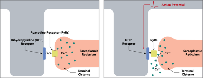
Cav1.1 is a voltage-dependent calcium channel found in the transverse tubule of muscles. In skeletal muscle it associates with the ryanodine receptor RyR1 of the sarcoplasmic reticulum via a mechanical linkage. It senses the voltage change caused by the end-plate potential from nervous stimulation and propagated by sodium channels as action potentials to the T-tubules. It was previously thought that when the muscle depolarises, the calcium channel opens, allowing calcium in and activating RyR1, which mediates much greater calcium release from the sarcoplasmic reticulum. This is the first part of the process of excitation-contraction coupling, which ultimately causes the muscle to contract. Calcium entry through Cav1.1 is not required in skeletal muscle, as it is in cardiac muscle; Cav1.1 undergoes a conformational change which allosterically activates RyR1.[2]
Clinical significance
In hypokalemic periodic paralysis (HOKPP), the voltage sensors in domains 2 and 4 of Cav1.1 are mutated (loss-of-function), reducing the availability of the channel to sense depolarisation, and therefore it cannot activate the ryanodine receptor as efficiently. As a result, the muscle cannot contract very well and the patient is paralysed. The condition is hypokalemic because a low extracellular potassium ion concentration will cause the muscle to repolarise to the resting potential more quickly, so any calcium conductance that does occur cannot be sustained. It becomes more difficult to reach the threshold at which the muscle can contract, and even if this is reached then the muscle is more prone to relaxing. Because of this, the severity would be reduced if potassium ion concentrations are maintained. In contrast, hyperkalemic periodic paralysis refers to gain-of-function mutations in sodium channels that maintain muscle depolarisation and therefore are aggravated by high potassium ion concentrations.[3]
The European Malignant Hyperthermia Group accepts two mutations in CACNA1S as diagnostic for malignant hyperthermia.[4]
Blockers
Cav1.1 is blocked by dihydropyridine.
See also
References
- ↑ 1.0 1.1 "Entrez Gene: CACNA1S calcium channel, voltage-dependent, L type, alpha 1S subunit". https://www.ncbi.nlm.nih.gov/sites/entrez?Db=gene&Cmd=ShowDetailView&TermToSearch=779.
- ↑ "Identification of a region of RyR1 that participates in allosteric coupling with the alpha(1S) (Ca(V)1.1) II-III loop". J. Biol. Chem. 277 (8): 6530–5. February 2002. doi:10.1074/jbc.M106471200. PMID 11726651.
- ↑ "Muscle channelopathies and critical points in functional and genetic studies". J. Clin. Invest. 115 (8): 2000–9. August 2005. doi:10.1172/JCI25525. PMID 16075040.
- ↑ "European Malignant Hyperthermia Group: Mutations in RYR1". https://emhg.org/nc/genetics/mutations-in-ryr1/.
Further reading
- "International Union of Pharmacology. XLVIII. Nomenclature and structure-function relationships of voltage-gated calcium channels". Pharmacol. Rev. 57 (4): 411–25. 2005. doi:10.1124/pr.57.4.5. PMID 16382099.
- "Specific phosphorylation of a COOH-terminal site on the full-length form of the alpha 1 subunit of the skeletal muscle calcium channel by cAMP-dependent protein kinase". J. Biol. Chem. 267 (23): 16100–5. 1992. doi:10.1016/S0021-9258(18)41972-2. PMID 1322891.
- "cAMP-dependent protein kinase rapidly phosphorylates serine- 687 of the skeletal muscle receptor for calcium channel blockers". J. Biol. Chem. 263 (30): 15325–9. 1988. doi:10.1016/S0021-9258(19)37591-X. PMID 2844809.
- "Primary structure of the receptor for calcium channel blockers from skeletal muscle". Nature 328 (6128): 313–8. 1987. doi:10.1038/328313a0. PMID 3037387. Bibcode: 1987Natur.328..313T.
- "Cloning of the human skeletal muscle alpha 1 subunit of the dihydropyridine-sensitive L-type calcium channel (CACNL1A3)". Genomics 24 (3): 608–9. 1994. doi:10.1006/geno.1994.1677. PMID 7713519.
- "Hypokalemic periodic paralysis and the dihydropyridine receptor (CACNL1A3): genotype/phenotype correlations for two predominant mutations and evidence for the absence of a founder effect in 16 caucasian families". Am. J. Hum. Genet. 56 (2): 374–80. 1995. PMID 7847370.
- "Mutation in DHP receptor alpha 1 subunit (CACLN1A3) gene in a Dutch family with hypokalaemic periodic paralysis". J. Med. Genet. 32 (1): 44–7. 1995. doi:10.1136/jmg.32.1.44. PMID 7897626.
- "Assignment of the human gene for the alpha 1 subunit of the skeletal muscle DHP-sensitive Ca2+ channel (CACNL1A3) to chromosome 1q31-q32". Genomics 15 (1): 107–12. 1993. doi:10.1006/geno.1993.1017. PMID 7916735.
- "A calcium channel mutation causing hypokalemic periodic paralysis". Hum. Mol. Genet. 3 (8): 1415–9. 1994. doi:10.1093/hmg/3.8.1415. PMID 7987325.
- "Dihydropyridine receptor mutations cause hypokalemic periodic paralysis". Cell 77 (6): 863–8. 1994. doi:10.1016/0092-8674(94)90135-X. PMID 8004673.
- "The gene coding for the alpha 1 subunit of the skeletal dihydropyridine receptor (Cchl1a3 = mdg) maps to mouse chromosome 1 and human 1q32". Mamm. Genome 4 (9): 499–503. 1993. doi:10.1007/BF00364784. PMID 8118099.
- "Refined localization of the alpha 1-subunit of the skeletal muscle L-type voltage-dependent calcium channel (CACNL1A3) to human chromosome 1q32 by in situ hybridization". Genomics 19 (3): 561–3. 1994. doi:10.1006/geno.1994.1106. PMID 8188298.
- "Exclusion of defects in the skeletal muscle specific regions of the DHPR alpha 1 subunit as frequent causes of malignant hyperthermia". J. Med. Genet. 32 (11): 913–4. 1995. doi:10.1136/jmg.32.11.913. PMID 8592342.
- "The structure of the gene encoding the human skeletal muscle alpha 1 subunit of the dihydropyridine-sensitive L-type calcium channel (CACNL1A3)". Genomics 31 (3): 392–4. 1996. doi:10.1006/geno.1996.0066. PMID 8838325.
- "A genome wide search for susceptibility loci in three European malignant hyperthermia pedigrees". Hum. Mol. Genet. 6 (6): 953–61. 1997. doi:10.1093/hmg/6.6.953. PMID 9175745.
- "Malignant-hyperthermia susceptibility is associated with a mutation of the alpha 1-subunit of the human dihydropyridine-sensitive L-type voltage-dependent calcium-channel receptor in skeletal muscle". Am. J. Hum. Genet. 60 (6): 1316–25. 1997. doi:10.1086/515454. PMID 9199552.
- "Sorcin associates with the pore-forming subunit of voltage-dependent L-type Ca2+ channels". J. Biol. Chem. 273 (30): 18930–5. 1998. doi:10.1074/jbc.273.30.18930. PMID 9668070.
- "Gating of the L-type Ca channel in human skeletal myotubes: an activation defect caused by the hypokalemic periodic paralysis mutation R528H". J. Neurosci. 18 (24): 10320–34. 1998. doi:10.1523/JNEUROSCI.18-24-10320.1998. PMID 9852570.
- "Multiple regions of RyR1 mediate functional and structural interactions with alpha(1S)-dihydropyridine receptors in skeletal muscle". Biophys. J. 83 (6): 3230–44. 2002. doi:10.1016/S0006-3495(02)75325-3. PMID 12496092. Bibcode: 2002BpJ....83.3230P.
- "Identification of new polymorphisms in the CACNA1S gene". Clin. Chem. Lab. Med. 41 (1): 20–2. 2003. doi:10.1515/CCLM.2003.004. PMID 12636044.
External links
- GeneReviews/NCBI/NIH/UW entry on Malignant Hyperthermia Susceptibility
- CACNA1S+protein,+human at the US National Library of Medicine Medical Subject Headings (MeSH)
This article incorporates text from the United States National Library of Medicine, which is in the public domain.
 |


