Biology:Cadherin-2
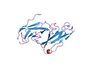
 Generic protein structure example |
Cadherin-2 also known as Neural cadherin (N-cadherin), is a protein that in humans is encoded by the CDH2 gene.[1][2][3] CDH2 has also been designated as CD325 (cluster of differentiation 325). Cadherin-2 is a transmembrane protein expressed in multiple tissues and functions to mediate cell–cell adhesion. In cardiac muscle, Cadherin-2 is an integral component in adherens junctions residing at intercalated discs, which function to mechanically and electrically couple adjacent cardiomyocytes. Alterations in expression and integrity of Cadherin-2 has been observed in various forms of disease, including human dilated cardiomyopathy. Variants in CDH2 have also been identified to cause a syndromic neurodevelopmental disorder.[4]
Structure
Cadherin-2 is a protein with molecular weight of 99.7 kDa, and 906 amino acids in length.[5] Cadherin-2, a classical cadherin from the cadherin superfamily, is composed of five extracellular cadherin repeats, a transmembrane region and a highly conserved cytoplasmic tail. Cadherin-2, as well as other cadherins, interact with Cadherin-2 on an adjacent cell in an anti-parallel conformation, thus creating a linear, adhesive "zipper" between cells.[6]
Function
Cadherin-2, originally named Neural cadherin for its role in neural tissue, plays a role in neurons and later was found to also play a role in cardiac muscle and in cancer metastasis. Cadherin-2 is a transmembrane, homophilic glycoprotein belonging to the calcium-dependent cell adhesion molecule family. These proteins have extracellular domains that mediate homophilic interactions between adjacent cells, and C-terminal, cytoplasmic tails that mediate binding to catenins, which in turn interact with the actin cytoskeleton.[7][8][9]
Role in development
Cadherin-2 plays a role in development as a calcium dependent cell–cell adhesion glycoprotein that functions during gastrulation and is required for establishment of left-right asymmetry.[10]
Cadherin-2 is widely expressed in the embryo post-implantation, showing high levels in the mesoderm with sustained expression through adulthood.[11] Cadherin-2 mutation during development has the most significant effect on cell adhesion in the primitive heart; dissociated myocytes and abnormal heart tube development occur.[12] Cadherin-2 plays a role in the development of the vertebrate heart at the transition of epithelial cells to trabecular and compact myocardial cell layer formation.[13] An additional study showed that myocytes expressing a dominant negative Cadherin-2 mutant showed significant abnormalities in myocyte distribution and migration towards the endocardium, resulting in defects in trabecular formation within the myocardium.[14][15]
Role in cardiac muscle
In cardiac muscle, Cadherin-2 is found at intercalated disc structures which provide end-on cell–cell connections that facilitate mechanical and electrical coupling between adjacent cardiomyocytes. Within intercalated discs are three types of junctions: adherens junctions, desmosomes and gap junctions;[16] Cadherin-2 is an essential component in adherens junctions, which enables cell–cell adhesion and force transmission across the sarcolemma.[17] Cadherin-2 complexed to catenins has been described as a master regulator of intercalated disc function.[18] Cadherin-2 appears at cell–cell junctions prior to gap junction formation,[19][20] and is critical for normal myofibrillogenesis.[21] Expression of a mutant form of Cadherin-2 harboring a large deletion in the extracellular domain inhibited the function of endogenous Cadherin-2 in adult ventricular cardiomyocytes, and neighboring cardiomyocytes lost cell–cell contact and gap junction plaques as well.[22]
Mouse models employing transgenesis have highlighted the function of N-cadherin in cardiac muscle. Mice with altered expression of N-cadherin and/or E-cadherin showed a dilated cardiomyopathy phenotype, likely due to malfunction of intercalated discs.[23] In agreement with this, mice with ablation of N-cadherin in adult hearts via a cardiac-specific tamoxifen-inducible Cre N-cadherin transgene showed disrupted assembly of intercalated discs, dilated cardiomyopathy, impaired cardiac function, decreased sarcomere length, increased Z-line thickness, decreases in connexin 43, and a loss in muscular tension. Mice died within two months of transgene expression, mainly due to spontaneous Ventricular tachycardia.[24] Further analysis of N-cadherin knockout mice revealed that the arrhythmias were likely due to ion channel remodeling and aberrant Kv1.5 channel function. These animals showed a prolonged action potential duration, reduced density of inward rectifier potassium channel and decreased expression of Kv1.5, KCNE2 and cortactin combined with disrupted actin cytoskeleton at the sarcolemma.[25]
Role in neurons
In neural cells, at certain central nervous system synapses, presynaptic to postsynaptic adhesion is mediated at least in part by Cadherin-2.[26] N-cadherins interact with catenins to play an important role in learning and memory (For full article see Cadherin-catenin complex in learning and memory). Loss of N-cadherin is also associated with attention-deficit hyperactivity disorder in humans, and impaired synaptic functioning. [27]
Role in cancer metastasis
Cadherin-2 is commonly found in cancer cells and provides a mechanism for transendothelial migration. When a cancer cell adheres to the endothelial cells of a blood vessel it up-regulates the src kinase pathway, which phosphorylates beta-catenins attached to both Cadherin-2 (this protein) and E-cadherins. This causes the intercellular connection between two adjacent endothelial cells to fail and allows the cancer cell to slip through.[28]
Clinical significance
Variants in CDH2 have been identified to cause a syndromic neurodevelopmental disorder characterized by Corpus callosum, axon, cardiac, ocular, and genital differences.[4]
One study investigating genetic underpinnings of obsessive-compulsive disorder and Tourette disorder found that while CDH2 variants are likely not disease-causing as single entities, they may confer risk when examined as part of a panel of related cell–cell adhesion genes.[29] Further studies in larger cohorts will be required to unequivocally determine this.
In human dilated cardiomyopathy, it was shown that Cadherin-2 expression was enhanced and arranged in a disarrayed fashion, suggesting that disorganization of Cadherin-2 protein in heart disease may be a component of remodeling.[30]
Interactions
Cadherin-2 has been shown to interact with:
- Beta-catenin,[31][32]
- CDH11,[31]
- type IIb RPTPs including PTPmu (CTNND1),[31][32]
- CTNNA1,[31][32]
- LRRC7,[33]
- PTPRM)[34][35]
- PTPrho (PTPRT),[36] and
- Plakoglobin.[31][37]
- XIRP1[38]
- SCARB2[39]
See also
References
- ↑ "UniProt". https://www.uniprot.org/uniprotkb/P19022/entry.
- ↑ "N-cadherin gene maps to human chromosome 18 and is not linked to the E-cadherin gene". Journal of Neurochemistry 55 (3): 805–12. September 1990. doi:10.1111/j.1471-4159.1990.tb04563.x. PMID 2384753.
- ↑ "Human N-cadherin: nucleotide and deduced amino acid sequence". Nucleic Acids Research 18 (19): 5896. October 1990. doi:10.1093/nar/18.19.5896. PMID 2216790.
- ↑ 4.0 4.1 "De Novo Pathogenic Variants in N-cadherin Cause a Syndromic Neurodevelopmental Disorder with Corpus Collosum, Axon, Cardiac, Ocular, and Genital Defects". American Journal of Human Genetics 105 (4): 854–868. October 2019. doi:10.1016/j.ajhg.2019.09.005. PMID 31585109.
- ↑ "Protein sequence of human CDH2 (Uniprot ID: P19022)". http://www.heartproteome.org/copa/ProteinInfo.aspx?QType=Protein%20ID&QValue=P19022.
- ↑ "Structural basis of cell-cell adhesion by cadherins". Nature 374 (6520): 327–37. March 1995. doi:10.1038/374327a0. PMID 7885471. Bibcode: 1995Natur.374..327S. http://orbit.dtu.dk/en/publications/structural-basis-of-cellcell-adhesion-by-cadherins(ec8eb34d-6db6-4bfb-8d7c-a5de4245c383).html.
- ↑ "Structure and interactions of desmosomal and other cadherins". Seminars in Cell Biology 3 (3): 157–67. June 1992. doi:10.1016/s1043-4682(10)80012-1. PMID 1623205.
- ↑ "Cadherins: a molecular family important in selective cell-cell adhesion". Annual Review of Biochemistry 59: 237–52. 1990. doi:10.1146/annurev.bi.59.070190.001321. PMID 2197976.
- ↑ "The cytoplasmic domain of the cell adhesion molecule uvomorulin associates with three independent proteins structurally related in different species". The EMBO Journal 8 (6): 1711–7. June 1989. doi:10.1002/j.1460-2075.1989.tb03563.x. PMID 2788574.
- ↑ "N-Cadherin, a cell adhesion molecule involved in establishment of embryonic left-right asymmetry". Science 288 (5468): 1047–51. May 2000. doi:10.1126/science.288.5468.1047. PMID 10807574. Bibcode: 2000Sci...288.1047G.
- ↑ "Dissociated spatial patterning of gap junctions and cell adhesion junctions during postnatal differentiation of ventricular myocardium". Circulation Research 80 (1): 88–94. January 1997. doi:10.1161/01.res.80.1.88. PMID 8978327.
- ↑ "Developmental defects in mouse embryos lacking N-cadherin". Developmental Biology 181 (1): 64–78. January 1997. doi:10.1006/dbio.1996.8443. PMID 9015265.
- ↑ "Differential adhesion leads to segregation and exclusion of N-cadherin-deficient cells in chimeric embryos". Developmental Biology 234 (1): 72–9. June 2001. doi:10.1006/dbio.2001.0250. PMID 11356020.
- ↑ "N-cadherin-catenin interaction: necessary component of cardiac cell compartmentalization during early vertebrate heart development". Developmental Biology 185 (2): 148–64. May 1997. doi:10.1006/dbio.1997.8570. PMID 9187080.
- ↑ "Trabecular myocytes of the embryonic heart require N-cadherin for migratory unit identity". Developmental Biology 193 (1): 1–9. January 1998. doi:10.1006/dbio.1997.8775. PMID 9466883.
- ↑ "Spatiotemporal relation between gap junctions and fascia adherens junctions during postnatal development of human ventricular myocardium". Circulation 90 (2): 713–25. August 1994. doi:10.1161/01.cir.90.2.713. PMID 8044940.
- ↑ "Intercalated discs of mammalian heart: a review of structure and function". Tissue & Cell 17 (5): 605–48. 1985. doi:10.1016/0040-8166(85)90001-1. PMID 3904080.
- ↑ "N-cadherin/catenin complex as a master regulator of intercalated disc function". Cell Communication & Adhesion 21 (3): 169–79. June 2014. doi:10.3109/15419061.2014.908853. PMID 24766605.
- ↑ "Dynamics of early contact formation in cultured adult rat cardiomyocytes studied by N-cadherin fused to green fluorescent protein". Journal of Molecular and Cellular Cardiology 32 (4): 539–55. April 2000. doi:10.1006/jmcc.1999.1086. PMID 10756112.
- ↑ "[Dynamic assembly of intercalated disc during postnatal development in the rat myocardium]". Sheng Li Xue Bao 66 (5): 569–74. October 2014. PMID 25332002.
- ↑ "The involvement of adherens junction components in myofibrillogenesis in cultured cardiac myocytes". Development 114 (1): 173–83. January 1992. doi:10.1242/dev.114.1.173. PMID 1576958.
- ↑ "N-cadherin in adult rat cardiomyocytes in culture. I. Functional role of N-cadherin and impairment of cell-cell contact by a truncated N-cadherin mutant". Journal of Cell Science 109 ( Pt 1) (1): 1–10. January 1996. doi:10.1242/jcs.109.1.1. PMID 8834785.
- ↑ "Remodeling the intercalated disc leads to cardiomyopathy in mice misexpressing cadherins in the heart". Journal of Cell Science 115 (Pt 8): 1623–34. April 2002. doi:10.1242/jcs.115.8.1623. PMID 11950881.
- ↑ "Induced deletion of the N-cadherin gene in the heart leads to dissolution of the intercalated disc structure". Circulation Research 96 (3): 346–54. February 2005. doi:10.1161/01.RES.0000156274.72390.2c. PMID 15662031.
- ↑ "Cortactin is required for N-cadherin regulation of Kv1.5 channel function". The Journal of Biological Chemistry 286 (23): 20478–89. June 2011. doi:10.1074/jbc.m111.218560. PMID 21507952.
- ↑ "Entrez Gene: CDH2 cadherin 2, type 1, N-cadherin (neuronal)". https://www.ncbi.nlm.nih.gov/sites/entrez?Db=gene&Cmd=ShowDetailView&TermToSearch=1000.
- ↑ "CDH2 mutation affecting N-cadherin function causes attention-deficit hyperactivity disorder in humans and mice". Nature Communications 6187 (12): 625–30. October 2021. doi:10.1038/s41467-021-26426-1. PMID 34702855.
- ↑ "Multi-scale modelling of cancer cell intravasation: the role of cadherins in metastasis". Physical Biology 6 (1): 016008. March 2009. doi:10.1088/1478-3975/6/1/016008. PMID 19321920. Bibcode: 2009PhBio...6a6008R.
- ↑ "Rare missense neuronal cadherin gene (CDH2) variants in specific obsessive-compulsive disorder and Tourette disorder phenotypes". European Journal of Human Genetics 21 (8): 850–4. August 2013. doi:10.1038/ejhg.2012.245. PMID 23321619.
- ↑ "Apoptosis-related factors p53, bcl-2 and the defects of force transmission in dilated cardiomyopathy". Pathology, Research and Practice 206 (9): 625–30. September 2010. doi:10.1016/j.prp.2010.05.007. PMID 20591580.
- ↑ 31.0 31.1 31.2 31.3 31.4 "A novel cell-cell junction system: the cortex adhaerens mosaic of lens fiber cells". Journal of Cell Science 116 (Pt 24): 4985–95. December 2003. doi:10.1242/jcs.00815. PMID 14625392.
- ↑ 32.0 32.1 32.2 "N-cadherin-catenin complexes form prior to cleavage of the proregion and transport to the plasma membrane". The Journal of Biological Chemistry 278 (19): 17269–76. May 2003. doi:10.1074/jbc.M211452200. PMID 12604612.
- ↑ "Densin-180 interacts with delta-catenin/neural plakophilin-related armadillo repeat protein at synapses". The Journal of Biological Chemistry 277 (7): 5345–50. February 2002. doi:10.1074/jbc.M110052200. PMID 11729199.
- ↑ "Receptor protein tyrosine phosphatase PTPmu associates with cadherins and catenins in vivo". The Journal of Cell Biology 130 (4): 977–86. August 1995. doi:10.1083/jcb.130.4.977. PMID 7642713.
- ↑ "Dynamic interaction of PTPmu with multiple cadherins in vivo". The Journal of Cell Biology 141 (1): 287–96. April 1998. doi:10.1083/jcb.141.1.287. PMID 9531566.
- ↑ "Intracellular substrates of brain-enriched receptor protein tyrosine phosphatase rho (RPTPrho/PTPRT)". Brain Research 1116 (1): 50–7. October 2006. doi:10.1016/j.brainres.2006.07.122. PMID 16973135.
- ↑ "Identification of plakoglobin domains required for association with N-cadherin and alpha-catenin". The Journal of Biological Chemistry 270 (34): 20201–6. August 1995. doi:10.1074/jbc.270.34.20201. PMID 7650039.
- ↑ "Localization of the novel Xin protein to the adherens junction complex in cardiac and skeletal muscle during development". Developmental Dynamics 225 (1): 1–13. September 2002. doi:10.1002/dvdy.10131. PMID 12203715.
- ↑ "Lysosomal integral membrane protein 2 is a novel component of the cardiac intercalated disc and vital for load-induced cardiac myocyte hypertrophy". The Journal of Experimental Medicine 204 (5): 1227–35. May 2007. doi:10.1084/jem.20070145. PMID 17485520.
Further reading
- "Shared cell adhesion molecule (CAM) homology domains point to CAMs signalling via FGF receptors". Perspectives on Developmental Neurobiology 4 (2–3): 157–68. 1997. PMID 9168198.
- "Follicular atresia and luteolysis. Evidence of a role for N-cadherin". Annals of the New York Academy of Sciences 900 (1): 46–55. 2000. doi:10.1111/j.1749-6632.2000.tb06215.x. PMID 10818391. Bibcode: 2000NYASA.900...46M.
- "Cadherin switch in tumor progression". Annals of the New York Academy of Sciences 1014 (1): 155–63. April 2004. doi:10.1196/annals.1294.016. PMID 15153430. Bibcode: 2004NYASA1014..155H.
- "N-cadherin as an invasion promoter: a novel target for antitumor therapy?". Current Opinion in Investigational Drugs 5 (12): 1274–8. December 2004. PMID 15648948.
- "Extrajunctional distribution of N-cadherin in cultured human endothelial cells". Journal of Cell Science 102 ( Pt 1) (1): 7–17. May 1992. doi:10.1242/jcs.102.1.7. PMID 1500442.
- "Plakoglobin, or an 83-kD homologue distinct from beta-catenin, interacts with E-cadherin and N-cadherin". The Journal of Cell Biology 118 (3): 671–9. August 1992. doi:10.1083/jcb.118.3.671. PMID 1639850.
- "Human N-cadherin: nucleotide and deduced amino acid sequence". Nucleic Acids Research 18 (19): 5896. October 1990. doi:10.1093/nar/18.19.5896. PMID 2216790.
- "N-cadherin gene maps to human chromosome 18 and is not linked to the E-cadherin gene". Journal of Neurochemistry 55 (3): 805–12. September 1990. doi:10.1111/j.1471-4159.1990.tb04563.x. PMID 2384753.
- "Expressed cadherin pseudogenes are localized to the critical region of the spinal muscular atrophy gene". Proceedings of the National Academy of Sciences of the United States of America 92 (9): 3702–6. April 1995. doi:10.1073/pnas.92.9.3702. PMID 7731968. Bibcode: 1995PNAS...92.3702S.
- "Structure of the human N-cadherin gene: YAC analysis and fine chromosomal mapping to 18q11.2". Genomics 22 (1): 172–9. July 1994. doi:10.1006/geno.1994.1358. PMID 7959764.
- "Expression and localization of N- and E-cadherin in the human testis and epididymis". International Journal of Andrology 17 (4): 174–80. August 1994. doi:10.1111/j.1365-2605.1994.tb01239.x. PMID 7995652.
- "Multiple cadherins are expressed in human fibroblasts". Biochemical and Biophysical Research Communications 235 (2): 355–8. June 1997. doi:10.1006/bbrc.1997.6707. PMID 9199196.
- "Differential localization of VE- and N-cadherins in human endothelial cells: VE-cadherin competes with N-cadherin for junctional localization". The Journal of Cell Biology 140 (6): 1475–84. March 1998. doi:10.1083/jcb.140.6.1475. PMID 9508779.
- "Distribution of N-cadherin and NCAM in neurons and endocrine cells of the human embryonic and fetal gastroenteropancreatic system". Acta Histochemica 100 (1): 83–97. February 1998. doi:10.1016/s0065-1281(98)80008-1. PMID 9542583.
- "Localization of human cadherin genes to chromosome regions exhibiting cancer-related loss of heterozygosity". Genomics 49 (3): 467–71. May 1998. doi:10.1006/geno.1998.5281. PMID 9615235.
- "delta-catenin, an adhesive junction-associated protein which promotes cell scattering". The Journal of Cell Biology 144 (3): 519–32. February 1999. doi:10.1083/jcb.144.3.519. PMID 9971746.
- "Functional cis-heterodimers of N- and R-cadherins". The Journal of Cell Biology 148 (3): 579–90. February 2000. doi:10.1083/jcb.148.3.579. PMID 10662782.
- "Proteomic analysis of NMDA receptor-adhesion protein signaling complexes". Nature Neuroscience 3 (7): 661–9. July 2000. doi:10.1038/76615. PMID 10862698.
External links
- CDH2+protein,+human at the US National Library of Medicine Medical Subject Headings (MeSH)
- CDH2 human gene location in the UCSC Genome Browser.
- CDH2 human gene details in the UCSC Genome Browser.
This article incorporates text from the United States National Library of Medicine, which is in the public domain.
 |


