Biology:CDH1 (gene)
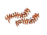
 Generic protein structure example |
Cadherin-1 (not to be confused with the APC/C activator protein CDH1) also known as CAM 120/80 or epithelial cadherin (E-cadherin) or uvomorulin is a protein that in humans is encoded by the CDH1 gene.[1] Mutations are correlated with gastric, breast, colorectal, thyroid, and ovarian cancers. CDH1 has also been designated as CD324 (cluster of differentiation 324). It is a tumor suppressor gene.[2][3]
Function
Cadherin-1 is a classical member of the cadherin superfamily. The encoded protein is a calcium-dependent cell–cell adhesion glycoprotein composed of five extracellular cadherin repeats, a transmembrane region, and a highly conserved cytoplasmic tail. Mutations in this gene are correlated with gastric, breast, colorectal, thyroid, and ovarian cancers. Loss of function is thought to contribute to progression in cancer by increasing proliferation, invasion, and/or metastasis. The ectodomain of this protein mediates bacterial adhesion to mammalian cells, and the cytoplasmic domain is required for internalization. Identified transcript variants arise from mutation at consensus splice sites.[4]
E-cadherin (epithelial) is the most well-studied member of the cadherin family. It consists of 5 cadherin repeats (EC1 ~ EC5) in the extracellular domain, one transmembrane domain, and an intracellular domain that binds p120-catenin and beta-catenin. The intracellular domain contains a highly-phosphorylated region vital to beta-catenin binding and, therefore, to E-cadherin function.[citation needed] Beta-catenin can also bind to alpha-catenin. Alpha-catenin participates in regulation of actin-containing cytoskeletal filaments. In epithelial cells, E-cadherin-containing cell-to-cell junctions are often adjacent to actin-containing filaments of the cytoskeleton.
E-cadherin is first expressed in the 2-cell stage of mammalian development, and becomes phosphorylated by the 8-cell stage, where it causes compaction.[citation needed] In adult tissues, E-cadherin is expressed in epithelial tissues, where it is constantly regenerated with a 5-hour half-life on the cell surface. [citation needed] Cell–cell interactions mediated by E-cadherin are crucial to blastula formation in many animals.[5]
Clinical significance

Loss of E-cadherin function or expression has been implicated in cancer progression and metastasis.[6][7] E-cadherin downregulation decreases the strength of cellular adhesion within a tissue, resulting in an increase in cellular motility. This in turn may allow cancer cells to cross the basement membrane and invade surrounding tissues.[7] E-cadherin is also used by pathologists to diagnose different kinds of breast cancer. When compared with invasive ductal carcinoma, E-cadherin expression is markedly reduced or absent in the great majority of invasive lobular carcinomas when studied by immunohistochemistry.[8]
Interactions
CDH1 (gene) has been shown to interact with
- CBLL1,[9]
- CDC27,[10]
- CDON,[11]
- CDH3,[12]
- C-Met,[13]
- CTNND1,[14][15][16]
- CTNNB1,[11][17][18][19]
- CTNNA1[20][21][22][23]
- FOXM1,[24]
- HDAC1,[25]
- HDAC2,[25]
- IQGAP1,[26]
- FYN,[27]
- NEDD9,[28]
- Plakoglobin,[17][29][30][31]
- Vinculin,[17][32]
Cancer
Metastasis
Transitions between epithelial and mesenchymal states play important roles in embryonic development and cancer metastasis. E-cadherin level changes in EMT (epithelial-mesenchymal transition) and MET (mesenchymal-epithelial transition). E-cadherin acts as an invasion suppressor and a classical tumor suppressor gene in pre-invasive lobular breast carcinoma.[36]
EMT
E-cadherin is a crucial type of cell–cell adhesion to hold the epithelial cells tight together. E-cadherin can sequester β-catenin on the cell membrane by the cytoplasmic tail of E-cadherin. Loss of E-cadherin expression results in releasing β-catenin into the cytoplasm. Liberated β-catenin molecules may migrate into the nucleus and trigger the expression of EMT-inducing transcription factors. Together with other mechanisms, such as constitutive RTK activation, E-cadherin loss can lead cancer cells to the mesenchymal state and undergo metastasis. E-cadherin is an important switch in EMT.[36]
MET
The mesenchymal state cancer cells migrate to new sites and may undergo METs in certain favorable microenvironment. For example, the cancer cells can recognize differentiated epithelial cell features in the new sites and upregulate E-cadherin expression. Those cancer cells can form cell–cell adhesions again and return to an epithelial state.[36]
Examples
- Inherited inactivating mutations in CDH1 are associated with Hereditary Diffuse Gastric Cancer. Individuals with this condition have up to a 70% lifetime risk of developing diffuse gastric carcinoma, and females with CDH1 mutations have up to a 60% lifetime risk of developing lobular breast cancer.[37]
- Inactivation of CDH1 (accompany with loss of the wild-type allele) in 56% of lobular breast carcinomas.[38][39]
- Inactivation of CDH1 in 50% of diffuse gastric carcinomas.[40]
- Complete loss of E-cadherin protein expression in 84% of lobular breast carcinomas.[41]
Genetic and epigenetic control
Several proteins such as SNAI1/SNAIL,[42][43] ZFHX1B/SIP1,[44] SNAI2/SLUG,[45][46] TWIST1[47] and DeltaEF1[48] have been found to downregulate E-cadherin expression. When expression of those transcription factors is altered, transcriptional repressors of E-cadherin were overexpressed in tumor cells.[42][43][44][45][47][48] Another group of genes, such as AML1, p300 and HNF3,[49] can upregulate the expression of E-cadherin.[50]
In order to study the epigenetic regulation of E-cadherin, M Lombaerts et al. performed a genome wide expression study on 27 human mammary cell lines. Their results revealed two main clusters that have the fibroblastic or epithelial phenotype, respectively. In close examination, the clusters showing fibroblast phenotypes only have either partial or complete CDH1 promoter methylation, while the clusters with epithelial phenotypes have both wild-type cell lines and cell lines with mutant CDH1 status. The authors also found that EMT can happen in breast cancer cell lines with hypermethylation of CDH1 promoter, but in breast cancer cell lines with a CDH1 mutational inactivation EMT cannot happen. It contradicts the hypothesis that E-cadherin loss is the initial or primary cause for EMT. In conclusion, the results suggest that “E-cadherin transcriptional inactivation is an epi-phenomenon and part of an entire program, with much more severe effects than loss of E-cadherin expression alone”.[50]
Other studies also show that epigenetic regulation of E-cadherin expression occurs during metastasis. The methylation patterns of the E-cadherin 5’ CpG island are not stable. During metastatic progression of many cases of epithelial tumors, a transient loss of E-cadherin is seen and the heterogeneous loss of E-cadherin expression results from a heterogeneous pattern of promoter region methylation of E-cadherin.[51]
See also
- Hereditary lobular breast cancer
- Cluster of differentiation
References
- ↑ "Assignment1 of the E-cadherin gene (CDH1) to chromosome 16q22.1 by radiation hybrid mapping". Cytogenetics and Cell Genetics 83 (1–2): 82–3. Mar 1999. doi:10.1159/000015134. PMID 9925936.
- ↑ "The tumor-suppressor function of E-cadherin". American Journal of Human Genetics 63 (6): 1588–93. December 1998. doi:10.1086/302173. PMID 9837810.
- ↑ "Adhesion-independent mechanism for suppression of tumor cell invasion by E-cadherin". The Journal of Cell Biology 161 (6): 1191–203. June 2003. doi:10.1083/jcb.200212033. PMID 12810698.
- ↑ "Entrez Gene: CDH1 cadherin 1, type 1, E-cadherin (epithelial)". https://www.ncbi.nlm.nih.gov/sites/entrez?Db=gene&Cmd=ShowDetailView&TermToSearch=999.
- ↑ "Assembly of tight junctions during early vertebrate development". Seminars in Cell & Developmental Biology 11 (4): 291–9. August 2000. doi:10.1006/scdb.2000.0179. PMID 10966863.
- ↑ "The E-cadherin-catenin complex in tumour metastasis: structure, function and regulation". European Journal of Cancer 36 (13 Spec No): 1607–20. August 2000. doi:10.1016/S0959-8049(00)00158-1. PMID 10959047.
- ↑ 7.0 7.1 Weinberg, Robert (2006). The Biology of Cancer. Garland Science. pp. 864 pages. ISBN 9780815340782. http://www.garlandscience.com/product/isbn/9780815340782. Retrieved 2012-05-06.
- ↑ Rosen, P. Rosen's Breast Pathology, 3rd ed, 2009, p. 704. Lippincott Williams & Wilkins.
- ↑ "Hakai, a c-Cbl-like protein, ubiquitinates and induces endocytosis of the E-cadherin complex". Nature Cell Biology 4 (3): 222–31. March 2002. doi:10.1038/ncb758. PMID 11836526.
- ↑ "TPR subunits of the anaphase-promoting complex mediate binding to the activator protein CDH1". Current Biology 13 (17): 1459–68. September 2003. doi:10.1016/S0960-9822(03)00581-5. PMID 12956947.
- ↑ 11.0 11.1 "Promyogenic members of the Ig and cadherin families associate to positively regulate differentiation". Proceedings of the National Academy of Sciences of the United States of America 100 (7): 3989–94. April 2003. doi:10.1073/pnas.0736565100. PMID 12634428. Bibcode: 2003PNAS..100.3989K.
- ↑ "Amino-terminal domain of classic cadherins determines the specificity of the adhesive interactions". Journal of Cell Science 113 (16): 2829–36. August 2000. doi:10.1242/jcs.113.16.2829. PMID 10910767.
- ↑ "HGF/SF modifies the interaction between its receptor c-Met, and the E-cadherin/catenin complex in prostate cancer cells". International Journal of Molecular Medicine 7 (4): 385–8. April 2001. doi:10.3892/ijmm.7.4.385. PMID 11254878.
- ↑ "The tyrosine kinase substrate p120cas binds directly to E-cadherin but not to the adenomatous polyposis coli protein or alpha-catenin". Molecular and Cellular Biology 15 (9): 4819–24. September 1995. doi:10.1128/mcb.15.9.4819. PMID 7651399.
- ↑ "A novel role for p120 catenin in E-cadherin function". The Journal of Cell Biology 159 (3): 465–76. November 2002. doi:10.1083/jcb.200205115. PMID 12427869.
- ↑ "Defining desmosomal plakophilin-3 interactions". The Journal of Cell Biology 161 (2): 403–16. April 2003. doi:10.1083/jcb.200303036. PMID 12707304.
- ↑ 17.0 17.1 17.2 "The epidermal growth factor receptor modulates the interaction of E-cadherin with the actin cytoskeleton". The Journal of Biological Chemistry 273 (15): 9078–84. April 1998. doi:10.1074/jbc.273.15.9078. PMID 9535896.
- ↑ "Expression and interaction of different catenins in colorectal carcinoma cells". International Journal of Molecular Medicine 8 (6): 695–8. December 2001. doi:10.3892/ijmm.8.6.695. PMID 11712088.
- ↑ "A truncated beta-catenin disrupts the interaction between E-cadherin and alpha-catenin: a cause of loss of intercellular adhesiveness in human cancer cell lines". Cancer Research 54 (23): 6282–7. December 1994. PMID 7954478.
- ↑ "Induction of tyrosine phosphorylation and association of beta-catenin with EGF receptor upon tryptic digestion of quiescent cells at confluence". Oncogene 15 (1): 71–8. July 1997. doi:10.1038/sj.onc.1201160. PMID 9233779.
- ↑ "Tyrosine phosphorylation regulates the adhesions of ras-transformed breast epithelia". The Journal of Cell Biology 130 (2): 461–71. July 1995. doi:10.1083/jcb.130.2.461. PMID 7542250.
- ↑ "Expression of E- or P-cadherin is not sufficient to modify the morphology and the tumorigenic behavior of murine spindle carcinoma cells. Possible involvement of plakoglobin". Journal of Cell Science 105 (4): 923–34. August 1993. doi:10.1242/jcs.105.4.923. PMID 8227214.
- ↑ "The fate of E- and P-cadherin during the early stages of apoptosis". Cell Death and Differentiation 6 (4): 377–86. April 1999. doi:10.1038/sj.cdd.4400504. PMID 10381631.
- ↑ "FoxM1 is degraded at mitotic exit in a Cdh1-dependent manner". Cell Cycle 7 (17): 2720–6. September 2008. doi:10.4161/cc.7.17.6580. PMID 18758239.
- ↑ 25.0 25.1 "WD repeat-containing mitotic checkpoint proteins act as transcriptional repressors during interphase". FEBS Letters 575 (1–3): 23–9. September 2004. doi:10.1016/j.febslet.2004.07.089. PMID 15388328.
- ↑ "IQGAP1 and calmodulin modulate E-cadherin function". The Journal of Biological Chemistry 274 (53): 37885–92. December 1999. doi:10.1074/jbc.274.53.37885. PMID 10608854.
- ↑ "p120 Catenin-associated Fer and Fyn tyrosine kinases regulate beta-catenin Tyr-142 phosphorylation and beta-catenin-alpha-catenin Interaction". Molecular and Cellular Biology 23 (7): 2287–97. April 2003. doi:10.1128/MCB.23.7.2287-2297.2003. PMID 12640114.
- ↑ "Direct interaction between Smad3, APC10, CDH1 and HEF1 in proteasomal degradation of HEF1". BMC Cell Biology 5 (1): 20. May 2004. doi:10.1186/1471-2121-5-20. PMID 15144564.
- ↑ "Association of plakoglobin with APC, a tumor suppressor gene product, and its regulation by tyrosine phosphorylation". Biochemical and Biophysical Research Communications 203 (1): 519–22. August 1994. doi:10.1006/bbrc.1994.2213. PMID 8074697.
- ↑ "Dynamics of cadherin/catenin complex formation: novel protein interactions and pathways of complex assembly". The Journal of Cell Biology 125 (6): 1327–40. June 1994. doi:10.1083/jcb.125.6.1327. PMID 8207061.
- ↑ "Plakoglobin, or an 83-kD homologue distinct from beta-catenin, interacts with E-cadherin and N-cadherin". The Journal of Cell Biology 118 (3): 671–9. August 1992. doi:10.1083/jcb.118.3.671. PMID 1639850.
- ↑ "Vinculin is associated with the E-cadherin adhesion complex". The Journal of Biological Chemistry 272 (51): 32448–53. December 1997. doi:10.1074/jbc.272.51.32448. PMID 9405455.
- ↑ "Receptor protein tyrosine phosphatase PTPmu associates with cadherins and catenins in vivo". The Journal of Cell Biology 130 (4): 977–86. August 1995. doi:10.1083/jcb.130.4.977. PMID 7642713.
- ↑ "Dynamic interaction of PTPmu with multiple cadherins in vivo". The Journal of Cell Biology 141 (1): 287–96. April 1998. doi:10.1083/jcb.141.1.287. PMID 9531566.
- ↑ "Intracellular substrates of brain-enriched receptor protein tyrosine phosphatase rho (RPTPrho/PTPRT)". Brain Research 1116 (1): 50–7. October 2006. doi:10.1016/j.brainres.2006.07.122. PMID 16973135.
- ↑ 36.0 36.1 36.2 "Transitions between epithelial and mesenchymal states: acquisition of malignant and stem cell traits". Nature Reviews. Cancer 9 (4): 265–73. April 2009. doi:10.1038/nrc2620. PMID 19262571.
- ↑ "Hereditary diffuse gastric cancer: updated clinical guidelines with an emphasis on germline CDH1 mutation carriers". Journal of Medical Genetics 52 (6): 361–74. June 2015. doi:10.1136/jmedgenet-2015-103094. PMID 25979631.
- ↑ "E-cadherin is a tumour/invasion suppressor gene mutated in human lobular breast cancers". The EMBO Journal 14 (24): 6107–15. December 1995. doi:10.1002/j.1460-2075.1995.tb00301.x. PMID 8557030.
- ↑ "E-cadherin is inactivated in a majority of invasive human lobular breast cancers by truncation mutations throughout its extracellular domain". Oncogene 13 (9): 1919–25. November 1996. PMID 8934538.
- ↑ "E-cadherin gene mutations provide clues to diffuse type gastric carcinomas". Cancer Research 54 (14): 3845–52. July 1994. PMID 8033105.
- ↑ "Simultaneous loss of E-cadherin and catenins in invasive lobular breast cancer and lobular carcinoma in situ". The Journal of Pathology 183 (4): 404–11. December 1997. doi:10.1002/(SICI)1096-9896(199712)183:4<404::AID-PATH1148>3.0.CO;2-9. PMID 9496256.
- ↑ 42.0 42.1 "The transcription factor snail is a repressor of E-cadherin gene expression in epithelial tumour cells". Nature Cell Biology 2 (2): 84–9. February 2000. doi:10.1038/35000034. PMID 10655587.
- ↑ 43.0 43.1 "The transcription factor snail controls epithelial-mesenchymal transitions by repressing E-cadherin expression". Nature Cell Biology 2 (2): 76–83. February 2000. doi:10.1038/35000025. PMID 10655586.
- ↑ 44.0 44.1 "The two-handed E box binding zinc finger protein SIP1 downregulates E-cadherin and induces invasion". Molecular Cell 7 (6): 1267–78. June 2001. doi:10.1016/S1097-2765(01)00260-X. PMID 11430829.
- ↑ 45.0 45.1 "The SLUG zinc-finger protein represses E-cadherin in breast cancer". Cancer Research 62 (6): 1613–8. March 2002. PMID 11912130.
- ↑ "The transcription factor snail induces tumor cell invasion through modulation of the epithelial cell differentiation program". Cancer Research 65 (14): 6237–44. July 2005. doi:10.1158/0008-5472.CAN-04-3545. PMID 16024625.
- ↑ 47.0 47.1 "Twist, a master regulator of morphogenesis, plays an essential role in tumor metastasis". Cell 117 (7): 927–39. June 2004. doi:10.1016/j.cell.2004.06.006. PMID 15210113.
- ↑ 48.0 48.1 "DeltaEF1 is a transcriptional repressor of E-cadherin and regulates epithelial plasticity in breast cancer cells". Oncogene 24 (14): 2375–85. March 2005. doi:10.1038/sj.onc.1208429. PMID 15674322.
- ↑ "Regulatory mechanisms controlling human E-cadherin gene expression". Oncogene 24 (56): 8277–90. December 2005. doi:10.1038/sj.onc.1208991. PMID 16116478.
- ↑ 50.0 50.1 "E-cadherin transcriptional downregulation by promoter methylation but not mutation is related to epithelial-to-mesenchymal transition in breast cancer cell lines". British Journal of Cancer 94 (5): 661–71. March 2006. doi:10.1038/sj.bjc.6602996. PMID 16495925.
- ↑ "Methylation patterns of the E-cadherin 5' CpG island are unstable and reflect the dynamic, heterogeneous loss of E-cadherin expression during metastatic progression". The Journal of Biological Chemistry 275 (4): 2727–32. January 2000. doi:10.1074/jbc.275.4.2727. PMID 10644736.
Further reading
- "Mutations of the human E-cadherin (CDH1) gene". Human Mutation 12 (4): 226–37. 1998. doi:10.1002/(SICI)1098-1004(1998)12:4<226::AID-HUMU2>3.0.CO;2-D. PMID 9744472.
- "E-cadherin-catenin cell–cell adhesion complex and human cancer". The British Journal of Surgery 87 (8): 992–1005. August 2000. doi:10.1046/j.1365-2168.2000.01513.x. PMID 10931041. http://repub.eur.nl/pub/56571.
- "The E-cadherin-catenin complex in tumour metastasis: structure, function and regulation". European Journal of Cancer 36 (13 Spec No): 1607–20. August 2000. doi:10.1016/S0959-8049(00)00158-1. PMID 10959047.
- "Polycystin: new aspects of structure, function, and regulation". Journal of the American Society of Nephrology 12 (4): 834–45. April 2001. doi:10.1681/ASN.V124834. PMID 11274246.
- "Germline E-cadherin gene mutations: is prophylactic total gastrectomy indicated?". Cancer 92 (1): 181–7. July 2001. doi:10.1002/1097-0142(20010701)92:1<181::AID-CNCR1307>3.0.CO;2-J. PMID 11443625.
- "Cadherin switch in tumor progression". Annals of the New York Academy of Sciences 1014 (1): 155–63. April 2004. doi:10.1196/annals.1294.016. PMID 15153430. Bibcode: 2004NYASA1014..155H.
- "The ins and outs of E-cadherin trafficking". Trends in Cell Biology 14 (8): 427–34. August 2004. doi:10.1016/j.tcb.2004.07.007. PMID 15308209.
- "CDH1 germline mutation in hereditary gastric carcinoma". World Journal of Gastroenterology 10 (21): 3088–93. November 2004. doi:10.3748/wjg.v10.i21.3088. PMID 15457549.
- "Regulation of cadherin stability and turnover by p120ctn: implications in disease and cancer". Seminars in Cell & Developmental Biology 15 (6): 657–63. December 2004. doi:10.1016/j.semcdb.2004.09.003. PMID 15561585.
- "CDH1 associated gastric cancer: a report of a family and review of the literature". European Journal of Surgical Oncology 31 (3): 259–64. April 2005. doi:10.1016/j.ejso.2004.12.010. PMID 15780560.
- "Role and expression patterns of E-cadherin in head and neck squamous cell carcinoma (HNSCC)". Journal of Experimental & Clinical Cancer Research 25 (1): 5–14. March 2006. PMID 16761612.
- "In the first extracellular domain of E-cadherin, heterophilic interactions, but not the conserved His-Ala-Val motif, are required for adhesion". The Journal of Biological Chemistry 277 (42): 39609–16. October 2002. doi:10.1074/jbc.M201256200. PMID 12154084.
External links
- CDH1+protein,+human at the US National Library of Medicine Medical Subject Headings (MeSH)
- GeneReviews/NCBI/NIH/UW entry on Hereditary Diffuse Gastric Cancer
- Human CDH1 genome location and CDH1 gene details page in the UCSC Genome Browser.
This article incorporates text from the United States National Library of Medicine, which is in the public domain.


